CHROMATOGRAPHY — Complete & Easy Notes
Theek hai, koi masla nahi! Ab main un saari 9 images se har topic cover karte hue ek complete, easy-words mein comprehensive note bana deta hoon — Chromatography ka pura concept, terminologies, theories, Paper
By Dr. Amina Rahman, PharmD13 min read
Reviewed by Medical Review Team ·
1. Definition & Introduction
Chromatography (Greek: chroma = color, graphein = to write) is a physical method of separation in which the components of a mixture are distributed between two phases:
- Stationary phase – stays fixed in one place
- Mobile phase – moves in a definite direction
The sample is dissolved in the mobile phase (gas, liquid, or supercritical fluid), which is then passed through an immobile stationary phase (solid or liquid). Because different components have different solubility/affinity for each phase, they travel at different speeds and get separated.
2. Important Terminologies
- Analyte – the substance being separated
- Chromatogram – the visual output/pattern of separation
- Chromatograph – the equipment used (e.g., GC, HPLC machine)
- Eluent – the solvent that carries the analyte (mobile phase going in)
- Eluate – the mobile phase coming out of the column (carrying separated substances)
- Bonded phase – stationary phase chemically attached to the support
- Immobilized phase – stationary phase fixed on the support particles
- Retention time – time taken by an analyte to pass through the system
- Solute – the sample components in partition chromatography
3. Adsorption vs Partition Chromatography (Comparison)
| Feature | Adsorption Chromatography | Partition Chromatography |
|---|---|---|
| Mechanism | Mobile phase gets adsorbed on the surface of solid stationary phase | Two immiscible liquids used; stationary phase is a liquid coated on solid support |
| Adsorbents/Support | Alumina, silica gel, calcium carbonate, starch, microcrystalline cellulose | Solvent coating on particles of solid support |
| Rate of separation | Depends on chemical nature & surface area of adsorbent + affinity of solute between two phases | Depends on solubility of solute — more soluble in mobile phase = more separation |
| Example | Column chromatography, TLC | Paper chromatography |
| Uses | Separating hydrocarbons/terpenes from oxygenated components; checking purity | Fractionation of biological mixtures, e.g., flavonoids from polyphenolic extract |
4. Theories of Chromatography
(a) Plate Theory
- Compares the column to fractional distillation (a theoretical/imaginary model).
- Assumes the column contains many separate layers called theoretical plates.
- Sample equilibrates between mobile and stationary phase at each plate.
- Rule: more theoretical plates = better separation.
- (Note: plates don't actually exist physically — it's just a way to measure efficiency, using "N" = number of theoretical plates, or HETP = Height Equivalent to a Theoretical Plate; smaller HETP = better.)
(b) Rate Theory
- The more realistic theory — depends on time of flow of mobile phase.
- Time of flow ∝ 1 / Rate of separation
- If flow time increases → rate of separation decreases → fewer/broader bands form.
(c) Band Broadening Theory
Explains why bands (spots) spread out instead of staying sharp. Three mechanisms:
- Eddy diffusion – solute molecules take random, different-length paths through the packed stationary phase, causing broadening.
- Longitudinal diffusion – analyte concentration is higher at the center of the band than at edges, so it diffuses outward. Faster mobile phase flow = less time for this = less broadening.
- Resistance to mass transfer – it takes time for analyte to equilibrate between stationary & mobile phase. If mobile phase flows too fast, equilibrium isn't reached properly, and the band broadens.
5. Plane Chromatography
This is a form of liquid chromatography where the stationary phase is on a flat/plane surface instead of inside a column. Common materials: glass, aluminium foil, plastic sheet coated with adsorbent.
Two main types:
- Paper Chromatography
- Thin Layer Chromatography (TLC)
6. Paper Chromatography (Detailed)
Definition: A method that separates and tests the purity of compounds using filter paper, based on the same principles as TLC — component distributes between a stationary phase (moisture held in paper) and a mobile phase (developing solvent moving up/along the paper).
Types of Paper Chromatography
(A) One-dimensional Separation
- Ascending – Paper hangs above a pool of solvent at the bottom; solvent rises up by capillary action.
- Descending – Solvent is in a trough above; it flows down the paper by capillary + gravity action together (faster separation).
(B) Two-dimensional Separation
- Sample is applied near a corner; developed once in one solvent, dried, rotated 90°, then developed again in a second (different) solvent. Good for complex mixtures.
(C) Radial (Circular) Separation
- Sample spotted at the center of a circular paper disc; a wick supplies solvent to the center, which then moves outward in circular paths.
Comparison Table:
| One-Dimensional | Two-Dimensional |
|---|---|
| One-way separation | Two-way separation |
| Separation in one dimension | Separation in two dimensions |
| Single direction/angle | Two directions/angles (90°) |
| Single run, single solvent | Two runs, two solvents |
| Simple process | Complex process |
Stationary Phase
- Usually Whatman filter paper (glass fiber paper, unaffected by reagents).
- Modified cellulose paper – used for amines, cations, amino acids (has carboxylic groups).
- Cellulose ester paper – used for most organic substances.
- Fiber orientation of the paper also affects how the mobile phase moves.
Mobile Phase
- Should be more polar than the paper's surface (since paper binds solute strongly).
- Common combination: n-butanol : water : acetic acid = 12 : 5 : 3
Procedure
- Prepare mobile phase (e.g., the butanol:water:acetic acid mixture above).
- Prepare the chamber – clean, dry, sealed developing tank; add ~2 cm mobile phase; let it saturate overnight.
- Prepare stationary phase (paper) – cut a square filter paper; draw a pencil line ~3 cm from the bottom (origin line).
- Spot the samples – apply with a micropipette, spot size 3–8 mm, spots 2–2.5 cm apart and away from edges.
- Develop the chromatogram – hang the paper in the chamber (don't touch the sides), dip bottom edge into solvent, let solvent rise/spread, then remove & dry in a well-ventilated area.
Identifying the Spots
- Visible spots → outline with pencil directly.
- Invisible spots, detected using:
- Chemical method – colourless compounds converted to coloured ones using a reagent
- Dipping technique – dip paper in reagent solution, dry.
- Spraying technique – spray reagent using atomizer, e.g., Ninhydrin for amino acids.
- Physical method
- UV detection – view under UV lamp (254 nm) — never look directly into the lamp.
- Radioactivity – detect radioactive-labelled compounds using a special counter.
- Chemical method – colourless compounds converted to coloured ones using a reagent
Detection Reagents Table
| Reagent | Colour of Spot | Detects |
|---|---|---|
| Ninhydrin | Pink/Purple | Amino acids & amines |
| Iodine vapours | Brown | General organic & unsaturated compounds |
| Antimony chloride | Various | Steroids, acyclic compounds, vitamins, carotenoids |
| Bromophenol blue | Yellow | Carboxylic acids |
Interpreting the Data — Rf Value
$$Rf = \frac{\text{Distance moved by substance}}{\text{Distance moved by solvent front}}$$
- Rf depends on temperature, solvent, and type of paper — so results should be compared using known standards spotted alongside unknowns.
- A pure sample shows only one spot; an impure sample shows two or more spots.
Applications
- Separation of amino acids and peptides (protein structure studies)
- Routine examination of urine/body fluids for sugars & amino acids
- Separation of purine bases & nucleotides (nucleic acid study)
- Separation of steroids
- Analysis of polymers
- Investigation of phenolic materials in plants
- Separation of alkaloids
- Detection of metals in soil/geological samples
- Separation of radioisotopically labelled compounds
Advantages & Disadvantages
- Advantage: Very sensitive — even tiny amounts of compound can be located after separation.
- Disadvantage: Cannot be used for volatile substances like hydrocarbons and volatile fatty acids.
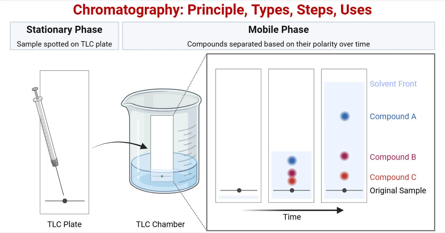
7. Thin Layer Chromatography (TLC) — Detailed
Definition: A method where a stationary phase (finely divided solid) is spread as a thin layer on a rigid plate, and the mobile phase (liquid) migrates across it.
Principle: Sample migrates up/across the stationary phase by capillary action of the mobile phase. Distance moved depends on the analyte's relative affinity/solubility for the stationary vs mobile phase.
Theory
- A substance strongly adsorbed → spends more time sitting still → moves less (stays near origin).
- A substance weakly adsorbed → spends more time moving with solvent → travels farther.
- Good separation needs adsorbent with high selectivity (a big difference in adsorption rate between substances).
Chromatographic Adsorbents (Order of Adsorption Strength)
| Most Strongly Adsorbent | Formula |
|---|---|
| Alumina | Al₂O₃ |
| Charcoal | C |
| Florisil | MgO/SiO₂ (anhydrous) |
| Least Strongly Adsorbent: Silica gel | SiO₂ |
Eluting Solvents (Eluotropic Series) — Alumina as adsorbent
From least eluting power to greatest eluting power:
Petroleum ether (hexane/pentane) → Cyclohexane → Carbon tetrachloride → Benzene → Dichloromethane → Chloroform → Ether (anhydrous) → Ethyl acetate (anhydrous) → Acetone (anhydrous) → Ethanol → Methanol → Water → Pyridine → Organic acids (greatest)
Stationary Phase
- Silica gel, alumina, or similar finely-ground material coated on a glass plate/metal/plastic film (~0.25 mm thick).
- A binder (like plaster of Paris/gypsum) helps it stick.
- Often mixed with a fluorescent indicator so spots can be seen under UV light (254 nm).
Mobile Phase — Criteria for Choosing a Solvent
- Solubility – must dissolve the sample well enough to move it up the plate.
- Affinity – sample shouldn't have too much affinity for the stationary phase (or it won't move — Rf too low).
- Resolution – optimizing affinity between sample, solvent & adsorbent to maximize separation between compounds.
Solvent Solubility Screening (increasing polarity):
Water → Methanol → Ethanol → Acetone → Diethyl Ether → Ethyl Acetate → Dichloromethane → Toluene → Chloroform → Cyclohexane → Petroleum Ether → Hexane
Affinity of Functional Groups for Silica Gel (increasing affinity):
Hydrocarbons → Halocarbons → Ether → Ester → Carbonyl → Amide → Alcohol → Carboxylic Acid → Amine
Procedure
- Preparation of plates – standard sizes: 5×20 cm, 10×20 cm, 20×20 cm (glass, plastic, stainless steel, or aluminium backing).
- Preparation of slurry – uniform-sized adsorbent particles mixed with water/organic solvent; a binder + fluorescent indicator may be added.
- Applying the slurry (3 methods):
- Spreading method – applicator/roller/glass rod spreads an even layer.
- Dipping method – used for microscope slides; dip glass slide into slurry, withdraw & dry.
- Tapping method – plastic tape applied to plate edges + center; slurry rolled on.
- Sample application – plates dried/conditioned; sample (few µg to mg) dissolved in a volatile solvent, spotted with a capillary tube or microliter syringe, keeping spots as small as possible.
- Detection methods – same as paper chromatography (chemical: dipping/spraying; physical: UV lamp, radioactivity/fluorescence quenching).
Fluorescence Quenching (special TLC technique): For colourless compounds — iodine vapours act as a "quencher," decreasing fluorescence at the spot location under UV light, making the compound visible against a glowing background.
Development Methods
- Radial separation – petri dish or jar; wick (dish) or thread (jar) carries solvent outward from center.
- Ascending development – plate stands in solvent; solvent flows upward via capillary action (most common).
- Descending development – solvent flows down; combines capillary + gravity, so separation is faster.
Rf Value (same formula as paper chromatography)
$$Rf = \frac{\text{Distance moved by substance}}{\text{Distance moved by solvent front}}$$
- Rf values depend strongly on adsorbent & solvent nature, so experimental Rf may not always match literature values exactly.
Applications
- Qualitative analysis (Rf values) & quantitative analysis (densitometry/scraping)
- Detection of narcotic/stimulant drugs
- Identification/isolation of active chemicals from plants & crude extracts
- Determination of glycerol in tobacco
- Determination of Vitamin B₁ in pharmaceutical products
- Determination of essential oils in herbal drugs
- Determination of plant pigments
- Detection of pesticides/insecticides in food
- Analysing dye composition of fibers (forensics)
- Monitoring chemical reactions & checking purity of reaction products
Importance of TLC (by field)
- Pharmaceutics & drugs – identification, purity testing, concentration of active ingredients
- Clinical/forensic/biochemistry – detecting metabolites, diagnosing metabolic disorders (e.g., phenylketonuria)
- Cosmetology – checking dyes, preservatives, surfactants, fatty acids in perfumes
- Food analysis – pesticide/fungicide residues, vitamins in soft drinks
- Environmental analysis – groundwater analysis, pollutant detection
Advantages of TLC over Paper Chromatography
- Faster runs
- Better separation
- Choice between different stationary phases
- Simplicity
- Speedy method
- No expensive equipment needed
8. Column Chromatography (Detailed)
Definition: A separation technique where a solid stationary phase (adsorbent) is held in a vertical glass tube (column); mobile phase is forced through it by gravity or pressure.
Types
- Gravity Column Chromatography – solvent flows down by gravity/percolation alone.
- Flash Chromatography – solvent is forced down using positive air pressure (pumps) — faster.
The Column
- Classical preparative column: glass tube, diameter 5 mm–50 mm, height 50 cm–1 m, with a tap and a filter (glass frit/wool plug) at the bottom to hold the stationary phase in.
Adsorbent (Stationary Phase)
- Must be solid, porous, finely divided powder.
- Most common: Silica gel, then Alumina, Cellulose Powder.
- Particle size matters:
- Smaller particles (e.g., 230–400 mesh) → used for Flash chromatography
- Larger particles (e.g., 70–230 mesh silica gel) → used for Gravity chromatography
Eluent (Mobile Phase)
- A pure solvent or mixture (e.g., hexane, ethyl acetate, ethanol, water).
- Polarity of solvent controls speed:
- Too polar → movement too rapid, poor separation.
- Polar enough (not appropriately chosen) → no compounds elute at all.
- Common strategy: start with a non-polar solvent (elutes less polar compounds first), gradually increase polarity to elute more polar compounds later.
Packing of the Column
- Prepare a slurry (adsorbent mixed with a solvent miscible with the eluent).
- Fix a cotton plug in the bottom of the glass column.
- Pour sand until a layer forms on the cotton.
- Add slurry on top of the sand layer, then place filter paper on the slurry.
- Add another sand layer on top (protects against disturbance while pouring solvent).
- Add mobile phase from the top.
- Open the stopper and let everything pack evenly/compress.
Sample Application
Sample is dissolved in a small amount of solvent (or as a concentrated solution) and added carefully to the top of the column.
Elution Procedure
- Mobile phase added from top; pinch clamp opened, allowed to drain to the top of the column bed.
- Small portions of solvent added and allowed to absorb into the adsorbent gradually; column filled to top with eluting solvent.
- Column run continuously, collecting fractions at the bottom as compounds elute.
- Compounds with more affinity for the stationary phase stay behind (retained longer).
- Solvent can be changed to more polar as needed to elute remaining compounds.
- Solvent level must never be allowed to drop below the top of the adsorbent bed — this would let air in and disturb the separation.
Analysis
- Coloured compounds – progress of separation can be visually monitored directly.
- Colourless compounds (more common) – small fractions collected at intervals in labelled test tubes, then each fraction analyzed by TLC — run on a TLC plate, spots identified under daylight or UV light. Each tube's separated component can then be identified.
Advantages
- Suitable for both preparative (isolating quantities) and analytical purposes.
- A pure single component can be separated out.
- Doesn't need very special/expensive equipment (compared to HPLC etc.)
Disadvantages
- More time-consuming compared to TLC or HPLC.
Ye ab in images mein maujood har single topic cover kar chuka hai — definitions, terminologies, theories, aur teeno major types (Paper, TLC, Column) — sab kuch simple English mein samjhaya hua hai.
Agar aap chahein to main ise ek Word (.docx) ya PDF file bana ke bhi de sakta hoon taake print/revise karna asan ho jaye — bata dein.
⚠️ Important Medical Disclaimer
This content is intended for educational and academic purposes only (e.g., Pharmacognosy/pharmacy coursework and exam preparation). It provides a general overview of chromatographic and spectroscopic techniques used in the analysis and quality control of natural and pharmaceutical compounds.
Related reading
If this article on CHROMATOGRAPHY — Complete & Easy Notes was helpful, explore these related guides for more practical information on similar topics.
- Extraction Methods of Alkaloids — Extraction Methods of Alkaloids: A Simple Pharm D Guide Introduction If you've studied pharmacognosy even a little, you already know alka…
- Solid Dosage Formulation Principles — Binders, disintegrants, and compression science for tablet development.
- Chemical Classification of Glycosides — Chemical Classification of Glycosides Introduction Glycosides are naturally occurring organic compounds in which a sugar molecule ( glyco…
- Plant-Derived Alkaloids: Is Kratom Itself an Alkaloid? — From isolation to therapeutic application — a structured review for pharmacy curricula.
- HPLC Method Validation in Pharmaceutical Analysis — ICH-aligned validation parameters for assay and impurity methods in QC laboratories.
- Theory and Applications of Atomic Absorption Spectroscopy (AAS) — Theory and Applications of Atomic Absorption Spectroscopy (AAS) Every time a lab checks whether a multivitamin actually has as much iron…