Apoptosis Defined: Mechanisms and Pharm-D Study Notes
Define apoptosis with clear pathways, morphology, clinical examples and exam distinctions, plus practical safety notes on apoptosis-targeting medicines.
By Dr. Amina Rahman, PharmD13 min read
Reviewed by Medical Review Team ·
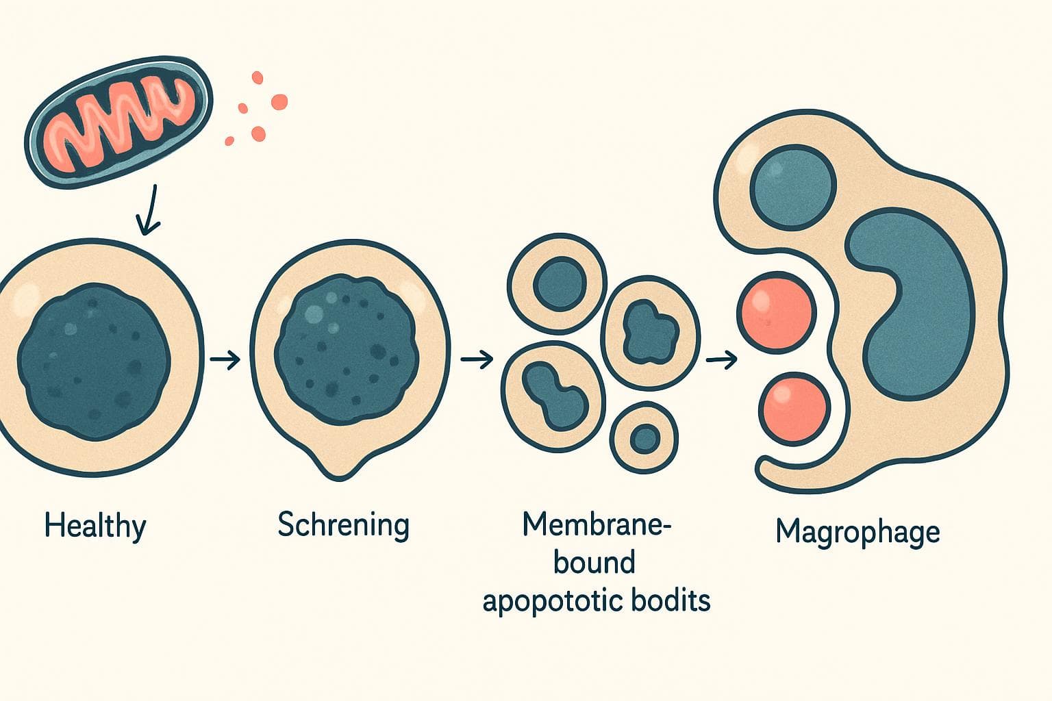
Apoptosis is a regulated process of cell death in which a cell activates molecular machinery to dismantle itself and allow its remains to be cleared, usually without significant inflammation. It removes unwanted, damaged or potentially harmful cells during normal development, tissue maintenance and disease.
These Pharm-D notes connect the definition to morphology, signaling pathways, laboratory interpretation and medicines. This is educational content, not advertising for any company's product; compare treatment options with a pharmacist rather than choosing by brand claims.
Quick Answer: What Makes Apoptosis Distinctive?
Apoptosis usually affects individual cells. The cell shrinks, its chromatin condenses, its DNA becomes fragmented, and membrane-bound pieces called apoptotic bodies may form. Neighboring cells or phagocytes then remove the remains.
The cleanup matters as much as the killing. When membranes remain intact until clearance, intracellular substances do not spill freely into surrounding tissue, explaining why uncomplicated apoptosis produces little inflammation compared with necrotic injury.
STICKY NOTE — Definition: Apoptosis is a form of regulated cell death, not a synonym for every form of cell death.
Key Definitions for Understanding the Pathway
- Caspases: Protein-cutting enzymes that use a cysteine residue at their active site and cleave substrates after aspartate residues. They drive the classical apoptotic pathway.
- Zymogen: An inactive enzyme precursor; many caspases initially exist as procaspases.
- Chromatin: DNA packaged with proteins inside the nucleus.
- BCL-2 family: Proteins that regulate the mitochondrial checkpoint, including both survival-promoting and death-promoting members.
- Cytochrome c: A mitochondrial electron-transfer protein that helps initiate apoptosis after entering the cytoplasm.
- Death receptor: A cell-surface receptor capable of transmitting a death signal under appropriate conditions.
- Efferocytosis: Recognition and removal of dying cells by phagocytes or neighboring cells.
Names can mislead students here. BCL-2 itself promotes survival, but other members of its protein family promote death; never assign function to the entire family.
When Apoptosis Occurs: Physiological and Pathological Examples
Physiological apoptosis
Normal tissues need controlled cell removal. During embryonic development, apoptosis helps shape structures, including the separation of developing digits. In adults, it supports turnover in continuously renewing tissues and contributes to hormone-dependent tissue regression.
Immune regulation supplies another clear example. Apoptosis removes many self-reactive lymphocytes during development and helps contract an expanded lymphocyte population after an immune response, although tolerance and immune resolution also require other mechanisms.
Pathological apoptosis
Cells may undergo apoptosis after irreparable DNA damage from radiation, cytotoxic medicines or other injuries. Accumulation of misfolded proteins can produce endoplasmic reticulum stress that activates apoptotic signaling if protective responses fail.
Some virus-infected cells are eliminated through apoptosis, including killing by cytotoxic lymphocytes. Obstruction-associated atrophy in glandular organs can also involve apoptotic loss.
Context determines the outcome. A similar insult may cause apoptosis at intensity and predominantly necrotic death at another, depending on the cell type, energy status and severity of injury.
Morphology: What Happens to the Dying Cell?
The classical sequence is recognizable:
- Cell shrinkage: The cytoplasm becomes denser, and the cell becomes smaller rather than swollen.
- Chromatin condensation: Nuclear material forms dense aggregates, often along the nuclear membrane.
- Nuclear fragmentation: The nucleus breaks into smaller fragments.
- Membrane blebbing: The cell surface develops outward bulges while initially retaining membrane integrity.
- Apoptotic body formation: Membrane-bound fragments contain portions of cytoplasm, organelles and nuclear material.
- Rapid engulfment: Nearby cells and macrophages remove the remnants.
Not every specimen shows every stage. Clearance can be so rapid that a tissue with substantial apoptotic activity contains relatively few visible apoptotic cells at any time.
On routine histology, apoptotic cells often appear as small, intensely eosinophilic structures with dense or fragmented nuclear material. Morphology remains valuable, but it should be interpreted alongside the tissue context.
Intrinsic Pathway: The Mitochondrial Checkpoint
The intrinsic pathway responds to internal stress. Common triggers include DNA damage, growth-factor withdrawal, severe protein-folding stress and certain metabolic disturbances.
Its central event is mitochondrial outer membrane permeabilization, often abbreviated MOMP. This allows proteins normally retained in the mitochondrial intermembrane space to enter the cytoplasm.
How the BCL-2 family controls commitment
Three functional groups are useful for exams:
| Group | Examples | Main role |
|---|---|---|
| Anti-apoptotic proteins | BCL-2, BCL-XL, MCL-1 | Restrain mitochondrial permeabilization |
| Pro-apoptotic effectors | BAX, BAK | Form membrane openings that permit protein release |
| BH3-only sensors | BIM, BID, BAD, PUMA, NOXA | Relay stress signals by countering survival proteins and/or activating effectors |
These proteins do not act as a simple numerical vote. Their binding partners, location, activation state and the cell's dependence on particular survival proteins influence whether death proceeds.
Following permeabilization, released cytochrome c binds apoptotic protease-activating factor 1, or Apaf-1. With nucleotide participation, this promotes assembly of the apoptosome, a protein platform that recruits and activates caspase-9.
Caspase-9 then activates executioner caspases, especially caspases-3 and -7. Mitochondrial proteins such as SMAC can also support apoptosis by opposing inhibitor-of-apoptosis proteins.
The tumor-suppressor protein p53 is relevant here. After DNA damage, it may promote repair or cell-cycle arrest; when damage cannot be resolved, it can favor apoptosis through targets including PUMA, NOXA and BAX. However, apoptosis does not always require p53.
Extrinsic Pathway: Signals from Outside the Cell
The extrinsic pathway begins with receptor signaling. A standard example is Fas ligand binding to the Fas receptor, also called CD95.
Activated Fas recruits an adaptor protein called Fas-associated death domain protein, or FADD. FADD helps assemble the death-inducing signaling complex, which recruits procaspase-8 and enables its activation.
Caspase-8 can activate executioner caspases directly. It can also cleave BID into truncated BID, which strengthens mitochondrial signaling; therefore, the extrinsic and intrinsic pathways are connected rather than completely separate routes.
Tumor necrosis factor receptor signaling requires caution in an exam answer. Depending on the signaling complex and cellular conditions, it may promote inflammation, survival, apoptosis or another regulated death pathway rather than automatically producing apoptosis.
Cytotoxic T cells and natural killer cells provide another route. They deliver granzymes with help from perforin; granzyme B can activate apoptotic machinery in a target cell without requiring the classical Fas receptor sequence.
Execution and Clearance: The Shared Final Steps
Executioner caspases cleave structural and regulatory proteins. They help dismantle the cytoskeleton and nuclear framework and permit activation of enzymes that fragment DNA.
For example, caspase cleavage of the inhibitor of caspase-activated DNase releases the DNase to cut chromosomal DNA. This explains the biochemical fragmentation associated with apoptosis, although a classic DNA ladder is not demonstrable in every setting.
The surface changes too. Phosphatidylserine, normally concentrated on the inner membrane leaflet, becomes exposed externally and helps mark the cell for removal.
Internal stress → BCL-2-family regulation → Mitochondrial permeabilization
 → Cytochrome c + Apaf-1 → Caspase-9
 ↓
Fas ligand → Fas/FADD signaling complex → Caspase-8 → Caspases-3/7
 ↓
 Cellular dismantling → Recognition → Clearance
REMEMBER — Low inflammation is conditional: If apoptotic cells are not cleared, they may lose membrane integrity and undergo secondary necrosis, allowing inflammation to develop.
Apoptosis Versus Necrosis: Important Differences
Do not reduce the distinction to “planned versus accidental.” Necrotic morphology can result from accidental injury or regulated pathways such as necroptosis.
| Feature | Typical apoptosis | Typical necrotic injury |
|---|---|---|
| Cell size | Shrinkage | Swelling |
| Plasma membrane | Preserved early; fragments remain membrane-bound | Integrity is lost |
| Nuclear changes | Condensation and fragmentation | Variable breakdown, including nuclear fading |
| Cell contents | Packaged for removal | Released into surrounding tissue |
| Inflammation | Usually limited with efficient clearance | Common, but context-dependent |
| Distribution | Often individual scattered cells | Often contiguous groups after shared injury |
| Molecular machinery | Classical forms use apoptotic caspases | Depends on the specific mechanism |
These are teaching patterns, not absolute diagnostic rules. Real lesions can contain several death mechanisms, and timing strongly affects what a biopsy or laboratory assay shows.
Also distinguish apoptosis from pyroptosis, a typically inflammatory, gasdermin-mediated death process, and ferroptosis, an iron-dependent process driven by lipid peroxidation.
How Apoptosis Is Studied: Avoid Single-Test Conclusions
No single result proves the whole mechanism. Researchers often combine morphology with membrane, enzyme and DNA measurements.
- Annexin V staining detects exposed phosphatidylserine. Pairing it with a membrane-impermeant dye helps distinguish populations with preserved versus disrupted membranes.
- Cleaved caspase-3 detection supports activation of executioner machinery, but the sampling time and experimental context matter.
- TUNEL testing detects DNA strand breaks. A positive result is not specific for apoptosis because other injuries and processes can generate detectable breaks.
- DNA fragmentation analysis may demonstrate an internucleosomal ladder, although its absence does not exclude apoptosis.
An important interpretation trap concerns late samples. Annexin V positivity together with uptake of a membrane-impermeant dye can reflect late apoptosis with secondary necrosis, but it cannot reliably exclude primary necrotic death by itself.
The practical lesson is straightforward: identify what the assay measures before naming the death pathway.
Clinical Significance and Pharmacy Practice
Too little or too much cell removal
Failure to eliminate damaged cells can contribute to cancer development and treatment resistance. BCL-2 overactivity is established survival mechanism; resistance can also involve altered death signaling, impaired drug exposure or other adaptations.
Excessive apoptotic loss can contribute to tissue injury, including neuronal and immune-cell depletion in certain diseases. Poor clearance of apoptotic material may also contribute to autoimmunity by increasing exposure to intracellular antigens.
Balance matters more than quantity. “More apoptosis” is not automatically beneficial, and a disease association does not prove that blocking apoptosis will improve clinical outcomes.
Medicines that influence apoptosis
Venetoclax, a selective BCL-2 inhibitor, is a direct clinical example. In susceptible malignant cells, it releases an important survival restraint and permits mitochondrial apoptotic signaling. Its approved uses and combinations depend on the jurisdiction and disease setting.
Cisplatin, a platinum antineoplastic medicine, causes DNA damage that can trigger apoptosis. Dexamethasone, a glucocorticoid, can promote apoptosis in susceptible lymphoid cells. Neither works exclusively through cell-death mechanism, and both have effects beyond apoptosis.
These are mechanistic examples, not interchangeable treatments. Apoptosis itself has no universal dose, pharmacokinetic profile or treatment schedule; those belong to each medicine and regimen.
A safety point students often miss
Orderly cell death can still produce a clinical emergency. Rapid destruction of a large tumor burden may cause tumor lysis syndrome, with dangerous changes in potassium, phosphate, calcium and uric acid that can injure the kidneys or disrupt heart rhythm.
Venetoclax therefore requires protocol-based risk assessment, dose escalation where indicated, preventive measures and laboratory monitoring. Other important risks include neutropenia and serious infection; interaction checks are essential because medicines affecting CYP3A or P-glycoprotein can change exposure.
Confirm the exact dose, schedule and any interruption or restart plan with the treating doctor or pharmacist and the current local product label. Never restart after a treatment gap from memory.
During cancer treatment, fever, new palpitations, confusion, markedly reduced urine or severe weakness needs urgent assessment. Do not simply stop and wait at home; contact the team urgently, or use emergency services for severe symptoms, and follow instructions about further doses.
IMPORTANT — Counseling insight: Little inflammation around an apoptotic cell does not mean that rapid tumor-cell death is harmless to the patient.
Common Mistakes and Confusions
Avoid these shortcuts:
- “Apoptosis happens in disease.” It is essential to normal development and tissue balance.
- “All caspases execute apoptosis.” Some have major inflammatory roles instead.
- “The membrane immediately ruptures.” Early preservation is a defining morphological feature.
- “Autophagy means apoptosis.” Autophagy recycles cellular material and often supports survival; the processes can interact but are distinct.
- “An apoptosis supplement treats cancer.” Laboratory findings do not establish safe, effective treatment in people.
Check the whole label. A supplement's claim to “activate cell death” is not evidence of selective tumor killing, and supplements can interact with prescribed cancer medicines.
Quick Pharm D Notes
- Definition: Regulated cellular dismantling with controlled removal of cellular remnants.
- Classification: Intrinsic mitochondrial and extrinsic death-receptor pathways are the classical routes.
- Initiators: Caspase-9 for the intrinsic pathway; caspase-8 for the classical extrinsic pathway.
- Executioners: Caspases-3 and -7 are central examples.
- Morphology: Shrinkage, condensed chromatin, fragmented nuclei and membrane-bound bodies.
- Examples: Embryonic tissue shaping, immune-cell selection and elimination after irreparable DNA injury.
- Clinical significance: Cancer resistance, tissue damage, immune regulation and drug response.
- Exam structure: Define apoptosis, list causes, describe morphology, explain both pathways and compare it with necrosis.
Frequently Asked Questions
Is apoptosis the same as programmed cell death?
Apoptosis is often called programmed cell death, especially in developmental contexts. The broader category of regulated cell death includes several non-apoptotic mechanisms, so the terms are not universally interchangeable.
Does apoptosis always require ATP?
Classical apoptosis is energy-dependent, and apoptosome assembly involves nucleotide binding. Severe energy depletion can change how an injured cell dies, but avoid presenting energy status as the deciding factor.
Why does apoptosis usually cause little inflammation?
Cell contents remain enclosed early, and phagocytes remove the dying cell promptly. Failed clearance or particular immune contexts can alter this pattern.
Which pathway involves cytochrome c?
The intrinsic mitochondrial pathway does. Released cytochrome c helps Apaf-1 assemble the apoptosome, leading to caspase-9 activation.
Can apoptosis be reversed?
Early stress signaling can resolve before commitment. Although recovery after some apoptotic events has been observed experimentally, established execution is generally treated as irreversible in routine teaching and clinical reasoning.
Can food or supplements safely increase apoptosis?
No general supplement strategy selectively increases apoptosis in harmful cells while protecting healthy tissue. Do not replace prescribed treatment with products marketed around laboratory cell-death findings.
Last-Minute Revision
- Apoptosis shrinks cells; necrotic injury typically swells them.
- BCL-2 restrains mitochondrial death, whereas BAX and BAK promote permeabilization.
- Cytochrome c, Apaf-1 and caspase-9 identify the intrinsic pathway.
- Fas, FADD and caspase-8 identify the classical extrinsic pathway.
- Executioner caspases dismantle cells; efferocytosis clears the remains.
- Interpret assays together, not in isolation.
References
- Robbins & Cotran Pathologic Basis of Disease: cell injury, cell death and adaptation.
- Alberts and colleagues, Molecular Biology of the Cell: apoptosis and cell-death regulation.
- Goodman & Gilman's The Pharmacological Basis of Therapeutics: antineoplastic pharmacology.
- DailyMed: current venetoclax prescribing information for indications, interactions and safety requirements.
Important Medical Disclaimer
This article is for general educational purposes and is not a substitute for advice from a qualified doctor or pharmacist. Always consult your doctor/pharmacist before using any medicine, changing a dose, or starting treatment.
Related reading
If this article on Apoptosis Defined: Mechanisms and Pharm-D Study Notes was helpful, explore these related guides for more practical information on similar topics.
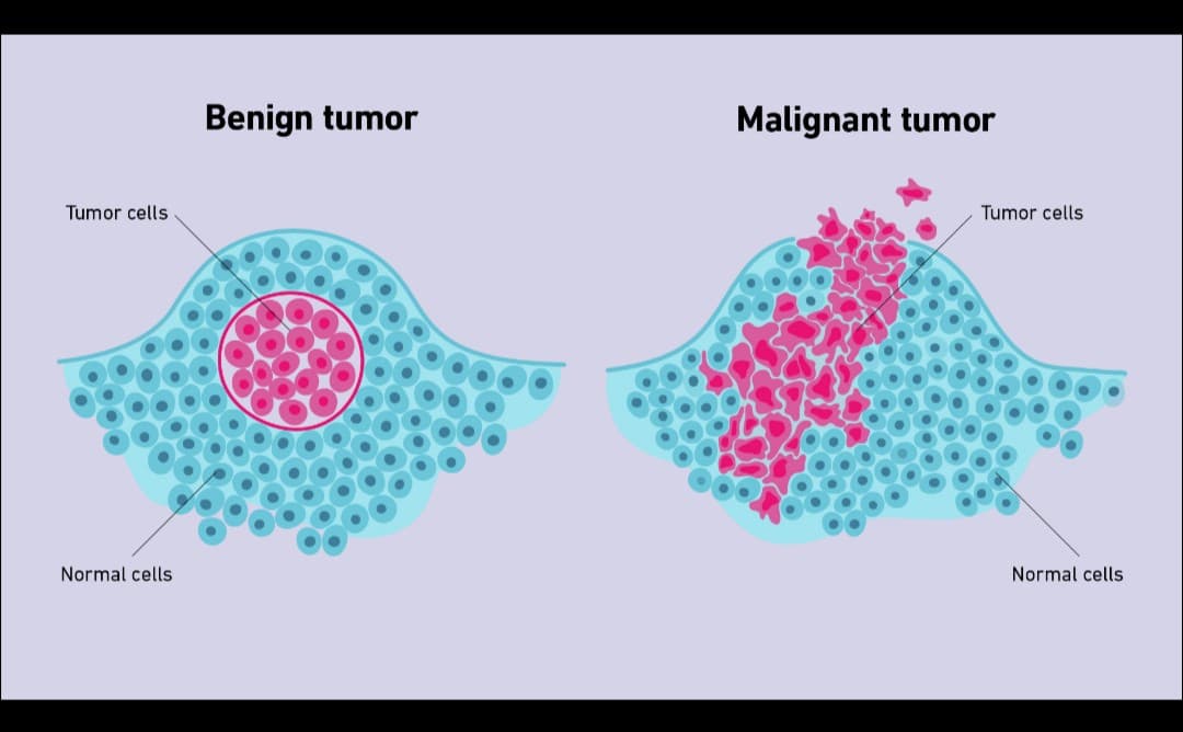
- Define neoplasia. Discuss in detail the features of malignant tumors. — Neoplasia — Definition Neoplasia is an abnormal, uncontrolled and excessive proliferation of cells that continues even after the original…
- Chronic Inflammation: Symptoms, Causes, Related Diseases & Treatment — Chronic Inflammation — Notes (Based on the Document) What is Chronic Inflammation? Chronic inflammation is a prolonged inflammatory respo…
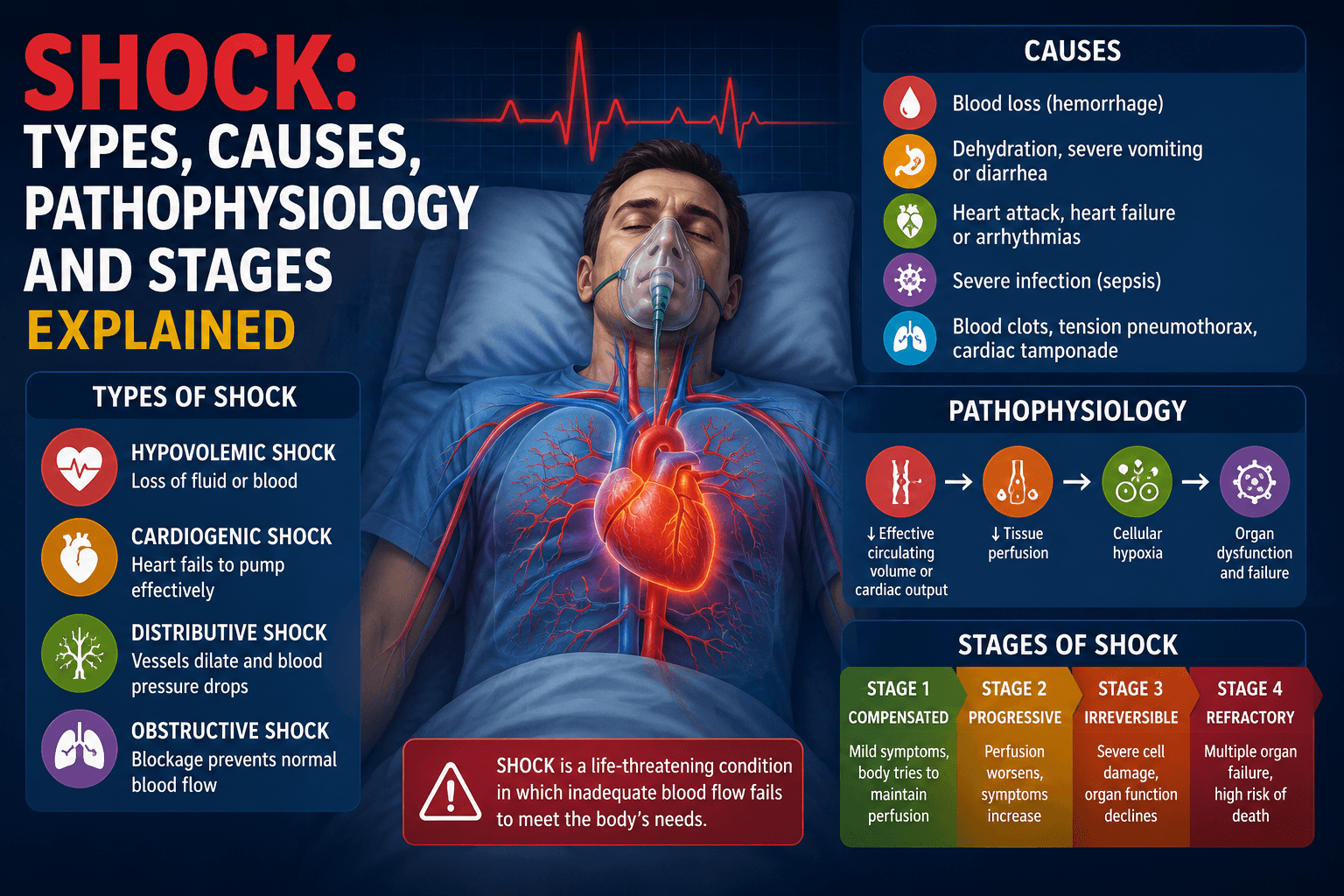
- Shock: Types, Causes, Pathophysiology and Stages Explained — Shock: Types, Causes, Pathophysiology and Stages Explained (Complete Pharm D Notes) Introduction Shock is of the most important topic…
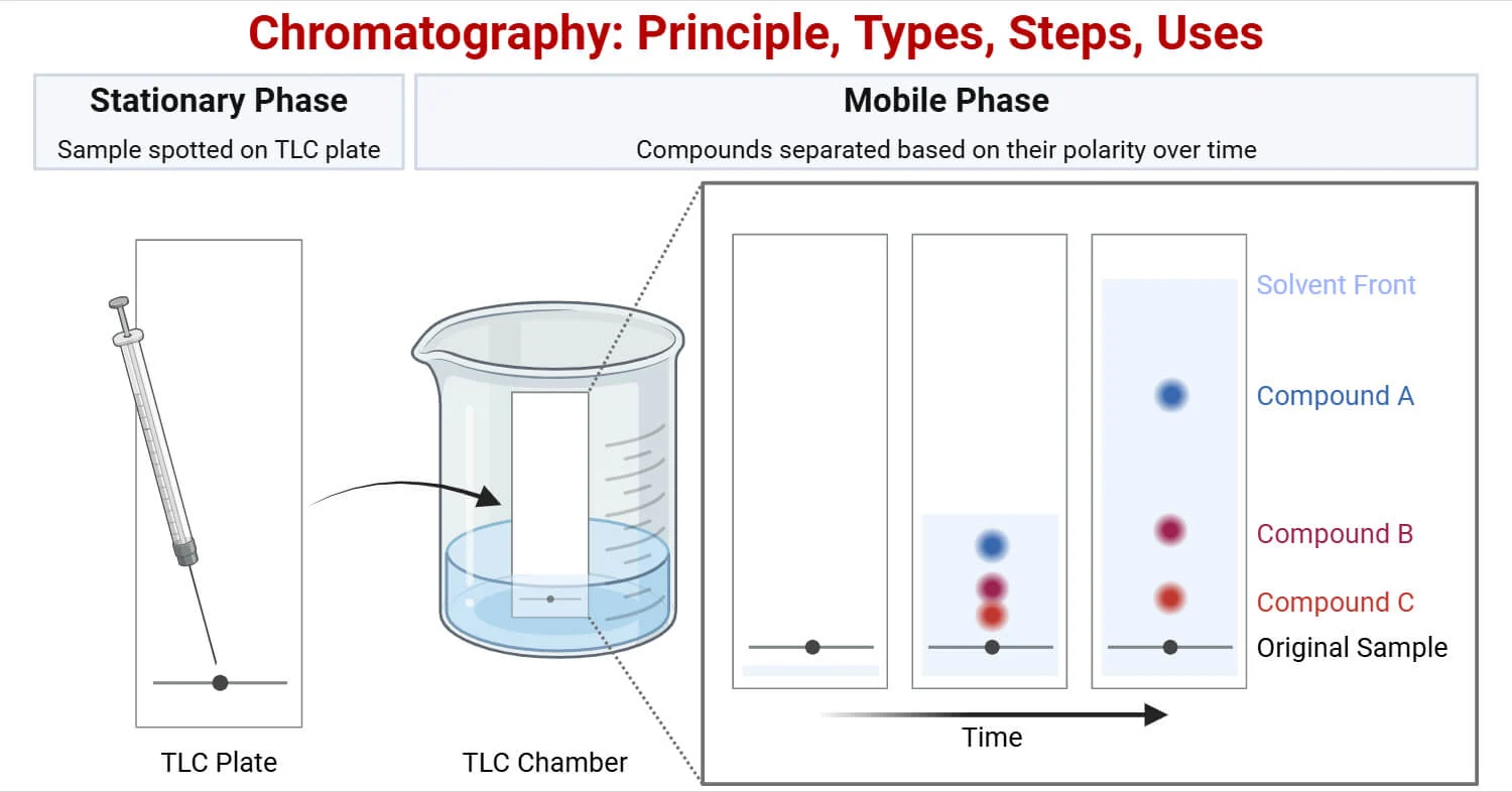
- CHROMATOGRAPHY — Complete & Easy Notes — Theek hai, koi masla nahi! Ab main un saari 9 images se har topic cover karte hue ek complete, easy-words mein comprehensive note bana de…
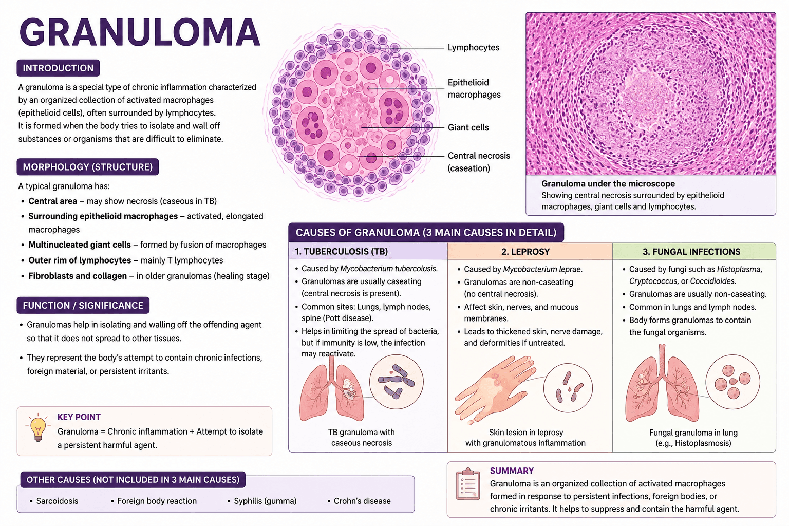
- write a note on granuloma and write down 3 causes of granuloma in details — How Patients Find the Right Doctor Today Finding the right doctor used to mean asking a neighbor, calling your insurance company,…
- Define acute inflammation describe different morphological patterns of acute inflammation — Definition Acute inflammation is a rapid and short-duration inflammatory response of vascularized tissue to injury or infection. It is ch…