Define neoplasia. Discuss in detail the features of malignant tumors.
Neoplasia — Definition Neoplasia is an abnormal, uncontrolled and excessive proliferation of cells that continues even after the original stimulus that caused the proliferation has been removed. The abnormal mass of
By Dr. Khalid Mehmood, MD11 min read
Neoplasia Explained: Definition, Benign vs Malignant Tumors, and the 10 Key Features of Malignancy
Introduction
Ask any Pharm D student what topic quietly underpins half of pathology and pharmacology, and neoplasia usually comes up. It's the reason chemotherapy drugs work the way they do, the reason a biopsy report reads the way it does, and honestly, it's of those topics that seems simple until an exam question hides an exception in it.
So let's build this up properly — starting with what neoplasia even means, then the difference between benign and malignant growths, and finally the ten features pathologists actually look for before they'll call something malignant.
Quick Answer / Overview
In plain terms, neoplasia is uncontrolled cell growth that doesn't stop — even after whatever triggered it in the first place is gone. The mass this produces is a neoplasm, which most people just call a tumor. Tumors come in two broad flavors: benign that stay put and mind their own business, and malignant that invade nearby tissue and can travel elsewhere in the body. Malignant is the we call cancer.
Key Definitions
- Neoplasia — abnormal, uncontrolled cell proliferation that keeps going even the original stimulus is removed.
- Neoplasm / Tumor — the physical mass this growth creates. ("Tumor" technically just meant "swelling" upon a time, but nobody uses it that way now — it's basically a synonym for neoplasm.)
- Cancer — reserved specifically for malignant neoplasms. You'll never hear a doctor call a benign lump "cancer."
- Benign — stays where it started; doesn't invade or spread.
- Malignant — invades surrounding tissue and can spread (metastasize).
- Differentiation — how much a tumor cell still resembles the mature, normal cell it came from.
- Anaplasia — when a tumor cell has lost that resemblance almost entirely and looks primitive.
- Metastasis — tumor cells setting up somewhere far from where they started.
What Is Neoplasia, Really?
The definition most textbooks lean on comes from the pathologist Rupert Willis: neoplasia is an abnormal mass of tissue whose growth exceeds, and is uncoordinated with, that of normal tissue — and it keeps growing that way even after whatever caused it in the first place is gone.
Read that last part again, because students tend to skip past it. A callus grows because of friction and stops the friction stops. A neoplasm doesn't play by that rule; it's grown past the point of needing permission.
Tumor vs. Neoplasm vs. Cancer
People use these three words like they mean the same thing, and mostly it doesn't matter — until it does. "Tumor" is the casual, everyday word for any abnormal mass. "Neoplasm" is the precise term pathology actually uses. "Cancer," though, is never casual — it's reserved strictly for malignant neoplasms, never benign ones.
📝 STICKY NOTE Every cancer is a neoplasm. Not every neoplasm is a cancer. Get that straight and half your terminology confusion disappears on its own.
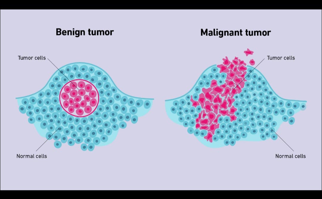
Benign vs. Malignant — Side by Side
| Feature | Benign | Malignant |
|---|---|---|
| Growth rate | Slow, usually | Fast |
| Differentiation | Well differentiated | Often poorly differentiated |
| Borders | Clean, often encapsulated | Ragged, poorly defined |
| Local invasion | Pushes tissue aside instead | Infiltrates and destroys it |
| Metastasis | Doesn't happen | Common (with a few exceptions) |
| Recurrence after removal | Rare | Common if not fully removed |
| Danger to the patient | Usually low, unless location is critical | Potentially life-threatening |
⚠️ IMPORTANT Don't equate "benign" with "harmless." A benign tumor sitting on the brainstem can kill a patient just as effectively as a malignant somewhere else — location matters as much as behavior does.
How These Tumors Get Named
Naming mostly follows the tissue of origin, and the suffix flips depending on whether the growth is benign or malignant:
| Tissue of Origin | Benign | Malignant |
|---|---|---|
| Glandular epithelium | Adenoma | Adenocarcinoma |
| Surface epithelium | Papilloma | Carcinoma |
| Fibrous tissue | Fibroma | Fibrosarcoma |
| Fat | Lipoma | Liposarcoma |
| Bone | Osteoma | Osteosarcoma |
| Smooth muscle | Leiomyoma | Leiomyosarcoma |
📝 STICKY NOTE Watch out for this — lymphoma, melanoma, seminoma, and mesothelioma are all malignant, despite ending in "-oma." These names got fixed into medical use before naming conventions were cleaned up, and they show up in exams precisely because they break the pattern.
The 10 Key Features of Malignant Tumors
Pathologists don't diagnose malignancy off single sign — it's a cluster of features that, taken together, tell the story of a cell that's stopped playing by the rules. Here's how that story usually unfolds.
It starts with growth that simply won't quit. Malignant cells keep dividing without waiting for the normal stop signal, so the mass keeps enlarging — and the faster it grows, the sooner symptoms tend to show up and the more urgent treatment becomes. Underneath that growth is a loss of identity: the cells stop resembling the tissue they came from, a change called anaplasia, or poor differentiation. This matters because poorly differentiated tumors are generally higher-grade and carry a worse outlook.
💡 REMEMBER Anaplasia doesn't mean a cell is going "backward" in development. It means the cell never finished becoming what it was supposed to become.
Look at these cells under a microscope and a few things jump out immediately. There's pleomorphism — real variation in cell and nuclear size and shape, even within the same tumor — which pathologists lean on heavily when grading how aggressive something looks. There's also abnormal mitosis: not just more cell division, but division that looks wrong, with multipolar or tripolar spindles instead of the usual clean split. And the nuclei themselves tend to be enlarged, dark-staining, and out of proportion to the surrounding cytoplasm — what's usually described as a high nuclear-to-cytoplasmic ratio. Put those three together and you've basically described what a pathologist is scanning for the moment a slide goes under the lens.
Then there's what happens at the tissue level rather than the cell level. Normal tissue has an orderly arrangement — cells know which way is "up." Malignant cells lose that orientation, a loss of architecture and polarity, and that disorganization is part of why the organ around it stops working properly. Worse, instead of staying contained, these tumors show local invasion — pushing into and destroying whatever's nearby, which is exactly what makes surgery complicated and what tends to cause organ-specific symptoms in the first place.
The most consequential feature, though, is metastasis — spread to lymph nodes or distant organs through lymphatics, blood vessels, or across body cavities. This single feature accounts for most cancer deaths and sits at the core of how cancers get staged.
⚠️ IMPORTANT If you remember exception for exams, make it this one: basal cell carcinoma is malignant and locally destructive, yet it almost never metastasizes. Metastasis is the most reliable sign of malignancy — it just isn't the requirement for it.
Two more features round this out. Tumors often outgrow their own blood supply, leaving patches of necrosis and hemorrhage — dead tissue and internal bleeding that can show up clinically as pain, fever, or ulceration. And to keep growing at all, tumors have to solve their own supply problem: they trigger angiogenesis, mostly through VEGF signaling, building new blood vessels toward themselves while reshaping the surrounding tissue and immune response to make room. This last isn't just academic — it's exactly why anti-angiogenic drugs like bevacizumab exist, and it directly influences how likely a tumor is to spread.
How Metastasis Actually Travels
                    MALIGNANT NEOPLASM
                           │
        ┌──────────────────┼──────────────────┐
        ↓                  ↓                  ↓
    Lymphatic          Hematogenous        Seeding across
   spread (to           spread (via         body cavities
   lymph nodes)       blood vessels —      (e.g. peritoneal
                     liver, lung, bone)     or pleural space)
        └──────────────────┼──────────────────┘
                           ↓
              Distant organ colonization
                    (metastasis)
📚 Quick Pharm D Notes
Definition: Uncontrolled cell proliferation that persists after the original stimulus is gone; produces a mass called a neoplasm.
Classification:
- Benign — localized, non-invasive, doesn't metastasize
- Malignant (cancer) — invasive, can metastasize
Underlying process:
- Escape from normal growth regulation
- Genomic instability → abnormal mitoses
- Loss of differentiation (anaplasia)
- Gained ability to invade and induce angiogenesis
Examples:
- Benign: lipoma, fibroma, adenoma, papilloma
- Malignant: adenocarcinoma, fibrosarcoma, lymphoma, melanoma
Clinical significance: Differentiation grade and metastatic spread drive both prognosis and treatment choice; angiogenesis is now a direct drug target.
Exam points worth memorizing:
- "-oma" isn't always benign — lymphoma, melanoma, seminoma, mesothelioma are the exceptions
- Metastasis is the most reliable sign of malignancy, but basal cell carcinoma rarely metastasizes despite being malignant
- Grading ≠ staging (grading = differentiation; staging = spread, via TNM)
- Anaplasia doesn't appear equally in every malignant tumor
A Memory Trick for Nuclear Features — "BIG HAND"
Just something to hang the histology details on:
- B — Big nucleus
- I — Irregular border
- G — Granular, coarse chromatin
- H — Hyperchromatic
- A — Abnormal mitoses
- N — N:C ratio increased
- D — Dark overall staining
(I made this up as a study aid — it's not from any textbook, just a way to hold the list together in your head.)
Where This Actually Matters in Practice
Pathologists use these features — differentiation, mitotic activity, pleomorphism — to grade a tumor, and that grade feeds straight into treatment decisions. On the drug side, a lot of chemotherapy is built around exactly these behaviors: vinca alkaloids and taxanes go after rapidly dividing cells, while anti-angiogenic agents target the VEGF pathway tumors depend on to build their own blood supply.
For pharmacists specifically, this knowledge shows up less in a lab and more in conversation — noticing when a patient's symptoms (a lump that's growing fast, unusual bleeding, sudden weight loss) warrant a referral rather than reassurance.
Common Mistakes and Confusions
- Assuming "tumor" automatically means cancer — it can be benign or malignant.
- Assuming every "-oma" is benign. Lymphoma, melanoma, seminoma, and mesothelioma say otherwise.
- Mixing up anaplasia, dysplasia, and metaplasia. They're related, but exams love testing exactly where they differ.
- Treating grading and staging as interchangeable when they measure completely different things.
- Assuming metastasis is mandatory for something to count as "malignant." Local invasion alone is enough — basal cell carcinoma proves it.
Terms That Get Confused With Each Other
| Term | What it means | Reversible? |
|---|---|---|
| Metaplasia | One mature cell type replaced by another mature type | Yes, if the trigger is removed |
| Dysplasia | Disordered growth, some loss of uniformity; often pre-neoplastic | Usually, if caught early |
| Anaplasia | Loss of differentiation in malignant cells | No |
| Neoplasia | Autonomous, uncontrolled proliferation forming a mass | No |
FAQ
Is every tumor cancerous? No. A tumor can be benign or malignant — "cancer" specifically means malignant.
Can a benign tumor become malignant later? Some can — certain colonic adenomas, for instance, can progress toward adenocarcinoma over time. It's not a rule that applies across the board, though.
What's the single best indicator that a tumor is malignant? Metastasis is usually treated as the most specific sign. Local invasion plus poor differentiation carries real weight too, especially since a few malignant tumors — basal cell carcinoma being the classic — almost never metastasize.
Why are lymphoma and melanoma malignant if they end in "-oma"? Mostly historical accident. These names were locked into medical usage before naming conventions got standardized, so students are generally just told to memorize the exceptions rather than derive them logically.
What actually drives angiogenesis in a tumor? Growth factors the tumor itself releases — VEGF being the main — which push nearby tissue to build new blood vessels toward it.
Is anaplasia basically the same as poor differentiation? Close enough for most purposes. Anaplasia is really just the more extreme end of poor differentiation.
How is grading different from staging? Grading looks at how abnormal the cells look under a microscope. Staging looks at how far the disease has actually spread through the body — size, node involvement, distant spread.
⚡ Last-Minute Revision
- Neoplasia = growth that won't stop, even the trigger is gone
- Neoplasm = the resulting mass; cancer = malignant neoplasm, specifically
- Benign: slow, well differentiated, encapsulated, doesn't invade or spread
- Malignant: fast, often anaplastic, invasive, can metastasize
- 10 hallmarks: rapid growth, anaplasia, pleomorphism, abnormal mitoses, hyperchromatic nuclei/high N:C ratio, loss of architecture, local invasion, metastasis, necrosis/hemorrhage, angiogenesis
- "-oma" exceptions: lymphoma, melanoma, seminoma, mesothelioma — all malignant
- Metastasis travels via lymphatics, blood, or body-cavity seeding
- Basal cell carcinoma: malignant, but almost never metastasizes — classic exception
- Grading = differentiation; staging = spread (TNM)
- Metaplasia and dysplasia can reverse; anaplasia and neoplasia don't
References
- Kumar, Abbas, Aster. Robbins & Cotran Pathologic Basis of Disease — Neoplasia chapter
- World Health Organization (WHO) Classification of Tumours
- National Cancer Institute (NCI) — cancer biology definitions
This article is for educational purposes and isn't a substitute for professional medical advice.
Related reading
If this article on Define neoplasia. Discuss in detail the features of malignant tumors. was helpful, explore these related guides for more practical information on similar topics.
- Chronic Inflammation: Symptoms, Causes, Related Diseases & Treatment — Chronic Inflammation — Notes (Based on the Document) What is Chronic Inflammation? Chronic inflammation is a prolonged inflammatory respo…
- Adverse Drug Reaction Reporting Systems — Pharmacovigilance fundamentals for students and practicing pharmacists.
- Define acute inflammation describe different morphological patterns of acute inflammation — Definition Acute inflammation is a rapid and short-duration inflammatory response of vascularized tissue to injury or infection. It is ch…
- write a note on granuloma and write down 3 causes of granuloma in details — How Patients Find the Right Doctor Today Finding the right doctor used to mean asking a neighbor, calling your insurance company,…
- The "7 Minutes After Death" Claim — What's Actually True? — The "7 Minutes After Death" Claim — What's Actually True? A few weeks ago a classmate sent me a video that had been going around — the on…
- Understanding Drug–Drug Interactions in Clinical Pharmacy Practice — A practical overview of clinically significant interactions.