Define acute inflammation describe different morphological patterns of acute inflammation
Definition Acute inflammation is a rapid and short-duration inflammatory response of vascularized tissue to injury or infection. It is characterized mainly by exudation of fluid and plasma proteins and accumulation of
By Dr. Amina Rahman, PharmD3 min read
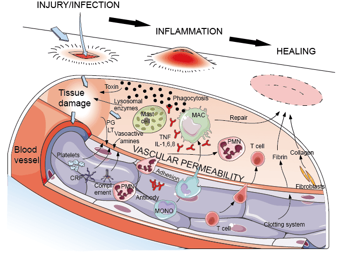
Definition
Acute inflammation is a rapid and short-duration inflammatory response of vascularized tissue to injury or infection. It is characterized mainly by exudation of fluid and plasma proteins and accumulation of neutrophils at the site of injury.
How you know it's happening (5 classic signs):
- 🔴 Redness
- 🔥 Heat
- 🫧 Swelling
- 😣 Pain
- ⚙️ Loss of function (the part doesn't work normally)
_(Radiopaedia_77488-89640_Longitudinal_1).jpeg/850px-Pseudomembranous_colitis_(gross_pathology)_(Radiopaedia_77488-89640_Longitudinal_1).jpeg)
Morphological Patterns of Acute Inflammation
Based on the severity of the reaction, causative agent, and the tissue/organ involved, acute inflammation can present in several distinct morphologic patterns:
1. Serous Inflammation
- Characterized by outpouring of thin, protein-poor fluid (derived from blood plasma or secretions of mesothelial cells).
- Example: Skin blister from a burn or viral infection; fluid in pleural, peritoneal, or pericardial cavities (effusion).
2. Fibrinous Inflammation
- Occurs when the vascular leak is severe enough to allow large molecules like fibrinogen to pass through and form fibrin.
- Typically develops on lining surfaces such as pericardium and pleura.
- Example: Fibrinous pericarditis ("bread and butter" appearance).
- May resolve completely (resolution) or organize into scar tissue if not removed (organization).
3. Suppurative (Purulent) Inflammation
- Characterized by production of pus — an exudate composed of neutrophils, necrotic cells, and edema fluid.
- Usually caused by pyogenic bacteria (e.g., Staphylococcus).
- Abscess: A localized collection of pus within a tissue, organ, or confined space, surrounded by inflamed tissue.
- Example: Acute appendicitis, boils, abscesses.
4. Ulcer
- A local defect (excavation) of the surface of an organ or tissue produced by sloughing (shedding) of inflamed necrotic tissue.
- Occurs when tissue necrosis and resulting inflammation exist on or near a surface.
- Example: Peptic ulcer, ulcers of the mouth or GI tract, skin ulcers in the lower extremities in the elderly.
5. Pseudomembranous Inflammation
- A special pattern occurring on mucosal surfaces (e.g., GI, respiratory tract) where a necrotizing inflammatory exudate combines with a superficial layer of fibrin and necrotic epithelial cells, forming a membrane-like layer ("pseudomembrane") over the mucosa.
- Example: Clostridioides difficile colitis (pseudomembranous colitis), diphtheria of the throat.
6. Catarrhal Inflammation
- Inflammation of a mucous membrane, characterized by hypersecretion of mucus.
- Example: Common cold (catarrhal rhinitis).
7. Hemorrhagic Inflammation
- Occurs when there is severe vascular damage leading to significant leakage of red blood cells along with the inflammatory exudate.
- Example: Severe viral hemorrhagic fevers, certain necrotizing infections.
Quick summary table:
Pattern Key Feature Example Serous Thin, protein-poor fluid Skin blister Fibrinous Fibrin-rich exudate Fibrinous pericarditis Suppurative/Purulent Pus formation Abscess, appendicitis Ulcer Surface tissue defect Peptic ulcer Pseudomembranous Necrotic mucosal membrane C. difficile colitis Catarrhal Excess mucus secretion Common cold Hemorrhagic RBC-rich exudate Hemorrhagic fever
Related reading
If this article on Define acute inflammation describe different morphological patterns of acute inflammation was helpful, explore these related guides for more practical information on similar topics.
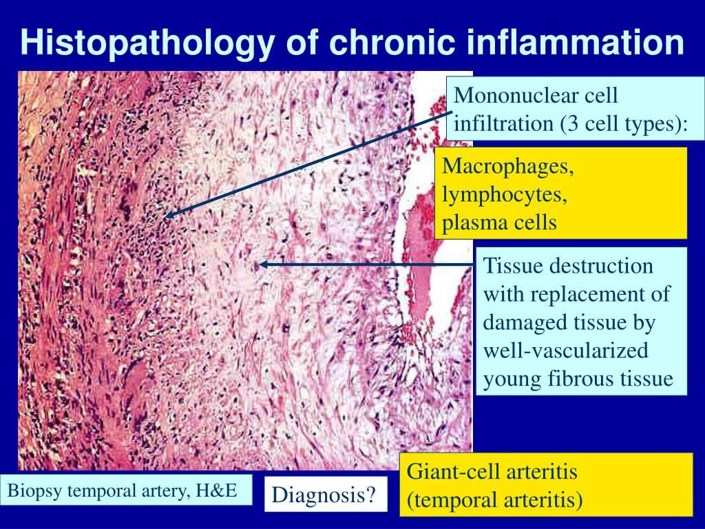
- Chronic Inflammation: Symptoms, Causes, Related Diseases & Treatment — Chronic Inflammation — Notes (Based on the Document) What is Chronic Inflammation? Chronic inflammation is a prolonged inflammatory respo…
- Understanding Drug–Drug Interactions in Clinical Pharmacy Practice — A practical overview of clinically significant interactions.
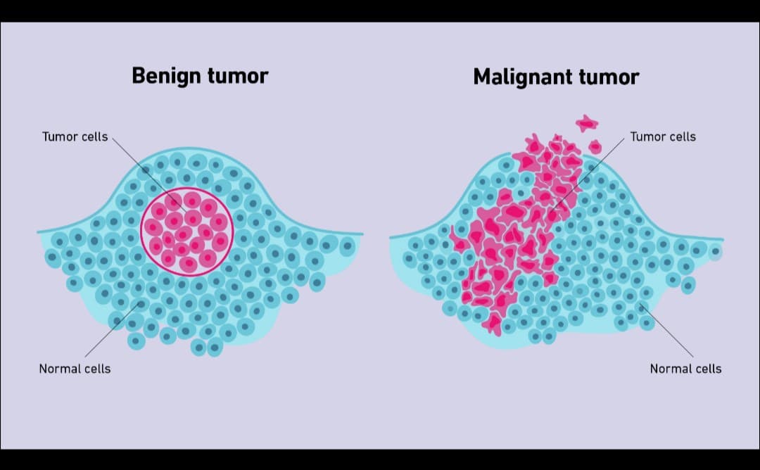
- Define neoplasia. Discuss in detail the features of malignant tumors. — Neoplasia — Definition Neoplasia is an abnormal, uncontrolled and excessive proliferation of cells that continues even after the original…
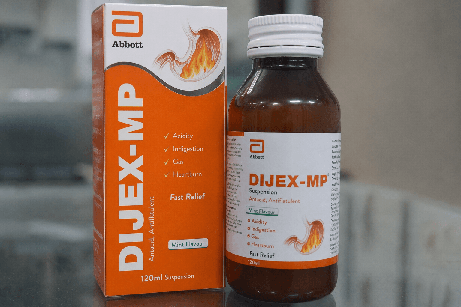
- dijex mp syrup uses in urdu — ڈائجکس ایم پی سیرپ (Dijex-MP) کے استعمالات — مکمل رہنما اور Pharm-D اسٹڈی نوٹس تعارف ڈائجکس ایم پی (Dijex-MP) پاکستان میں استعمال ہونے وا…
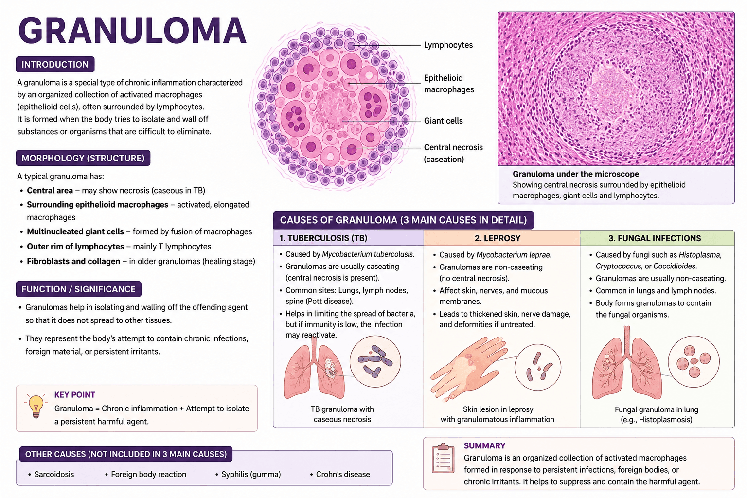
- write a note on granuloma and write down 3 causes of granuloma in details — How Patients Find the Right Doctor Today Finding the right doctor used to mean asking a neighbor, calling your insurance company,…
- Medication Therapy Management in Community Practice — How structured MTM visits improve outcomes and expand the pharmacist's clinical role.