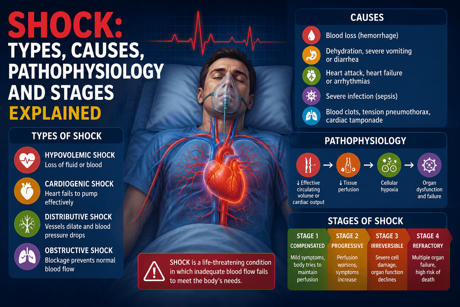
Shock: Types, Causes, Pathophysiology and Stages Explained
Shock: Types, Causes, Pathophysiology and Stages Explained (Complete Pharm D Notes) Introduction Shock is one of the most important topics in pathophysiology, and every Pharm D student runs into it early — in pathology,
By Dr. Amina Rahman, PharmD24 min read
Introduction
Shock is of the most important topics in pathophysiology, and every Pharm D student runs into it early — in pathology, physiology, and later in clinical pharmacy and critical care. In simple words, shock is not just "low blood pressure." It is a state where the circulatory system fails to deliver enough oxygen and nutrients to body tissues, and this failure can happen for very different reasons — from massive bleeding to a failing heart to a severely infected body.
Understanding shock properly means understanding why it happens (the cause), how the body tries to fight back (compensation), and what happens when the body loses that fight (progression to organ damage). This article walks through all of that in a simple, structured way, using the classic "container vs content" concept to make the four major types of shock easy to visualize and remember.
This article is written for educational purposes to help Pharm D students and healthcare learners understand the disease process. It is not a guide for diagnosing or treating any patient.
Quick Answer / Overview
Shock is a life-threatening condition in which blood flow to the tissues becomes so low that cells cannot get enough oxygen (hypoxia), and if not corrected, this leads to cell injury, organ failure, and death.
There are four major types of shock, based on what goes wrong:
| Type | What goes wrong |
|---|---|
| Hypovolemic | Not enough blood volume in the vessels |
| Cardiogenic | The heart fails as a pump |
| Distributive | Blood vessels dilate too much (container becomes too big) |
| Obstructive | Something physically blocks blood flow |
Shock develops in stages — starting with a stage the body can compensate for, moving to a stage where things start to break down, and finally (if untreated) reaching a stage that cannot be reversed even with treatment.
Key Definitions
- Shock: A clinical state of inadequate tissue perfusion (blood flow) relative to the metabolic needs of the tissue, resulting in cellular hypoxia (lack of oxygen at the cell level).
- Perfusion: The flow of blood through tissues, delivering oxygen and nutrients and removing waste.
- Hypoxia: A condition where tissues do not receive enough oxygen to function normally.
- Cardiac Output (CO): The amount of blood the heart pumps per minute. CO = Heart Rate (HR) × Stroke Volume (SV).
- Stroke Volume (SV): The volume of blood ejected by the left ventricle in heartbeat.
- Total Peripheral Resistance (TPR) / Systemic Vascular Resistance (SVR): The resistance blood faces as it flows through the vessels, mainly determined by the diameter of small arteries and arterioles.
- Preload: The degree of stretch on the ventricle before it contracts, largely determined by venous return.
- Vasoconstriction / Vasodilation: Narrowing / widening of blood vessels.
- Compensatory mechanism: A body response that tries to restore normal blood pressure and perfusion when they start to fall.
📝 STICKY NOTE Important point: Shock is defined by poor tissue perfusion, not simply by a low blood pressure number. In the early stage of shock, blood pressure can still look normal because the body is compensating hard behind the scenes.
Main Explanation
1. What Is Shock, Really?
At its core, shock happens when there is a mismatch between:
- The amount of blood the circulatory system needs to move (to keep tissues oxygenated), and
- Its actual ability to move that blood.
This mismatch can happen because there isn't enough blood (volume problem), the pump is broken (heart problem), the pipes have become too wide (vessel problem), or the pipes are blocked (obstruction problem). This is exactly the logic used to classify shock into four types.
2. The "Container vs Content" Way to Understand Shock
A simple and very effective way to understand the four types of shock is to think of the circulatory system as a container (the blood vessels) holding a content (the blood volume).
For normal circulation, the container's capacity and the volume of blood inside it must match reasonably well, and the pump (heart) must be working, and the pipe must be open (not obstructed).
- Hypovolemic shock: The container size is normal, but the content (volume) is reduced. (e.g., capacity ~5 L, but actual volume ~3 L)
- Cardiogenic shock: The container size and content volume are both normal, but the pump (heart) fails to push the content around effectively.
- Distributive shock: The content (volume) may be normal, but the container becomes far too large because the vessels dilate excessively — so the same amount of blood is now "too little" for the oversized container. (e.g., capacity increases to ~15 L, while volume stays ~5 L)
- Obstructive shock: The container and content are both normal, but something blocks the lumen (the pathway), so blood cannot flow through properly.
💡 REMEMBER Think of it like a bathtub: hypovolemic shock is not enough water, cardiogenic shock is a broken tap that won't push water out, distributive shock is the tub becoming too wide for the water in it, and obstructive shock is a blocked drain/pipe.
3. Classification of Shock — Overview Table
| Type of Shock | Core Problem | Typical Triggers |
|---|---|---|
| Hypovolemic | ↓ Circulating blood volume | Bleeding, fluid/plasma loss |
| Cardiogenic | Pump (heart) failure | MI, arrhythmia, cardiomyopathy, valve rupture |
| Distributive | Excessive vasodilation (container too big) | Sepsis, anaphylaxis, neurogenic causes |
| Obstructive | Physical obstruction to blood flow | Tamponade, tension pneumothorax, massive PE |
4. Hypovolemic Shock
Hypovolemic shock occurs when circulating blood volume drops significantly. The causes fall into two broad groups: loss of whole blood and loss of plasma/fluid.
A. Loss of Blood (Bleeding / Hemorrhage)
- External bleeding
- Trauma
- GIT (gastrointestinal) bleeding
- Internal bleeding
- Hemothorax (blood collecting in the pleural/chest cavity)
- Hemoperitoneum (blood collecting in the abdominal cavity) — a classic example is rupture of an ectopic pregnancy
B. Loss of Plasma or Fluid (Non-Hemorrhagic)
Plasma loss:
- Burns — large surface-area burns cause a huge amount of plasma to leak out of damaged capillaries into the tissues
- Exfoliative dermatitis — a severe skin condition/infection where large areas of skin are damaged, causing plasma loss through the skin
Fluid and electrolyte loss — External:
- Vomiting
- Diarrhea
- Polyuria (excess urination), such as in diabetic ketoacidosis (DKA)
- Excessive sweating
Fluid and electrolyte loss — Internal (third-spacing): This happens when fluid is not actually lost from the body, but gets trapped in a body compartment and is therefore "lost" from active circulation.
- Ascites (fluid accumulation in the abdominal cavity)
- Bowel obstruction
- Acute pancreatitis — when the pancreas becomes inflamed, the surrounding tissue also becomes inflamed, and fluid gets trapped in this inflamed region instead of staying in circulation
⚠️ IMPORTANT Third-spacing conditions (ascites, bowel obstruction, pancreatitis) cause hypovolemic shock without any visible external fluid loss — this is a commonly missed concept by students who think of hypovolemic shock as "bleeding" or "vomiting/diarrhea."
5. Cardiogenic Shock
Cardiogenic shock happens when the heart itself fails as a pump, even though blood volume is normal. The main causes include arrhythmias, pump (muscle) failure, valvular dysfunction, and mechanical rupture.
A. Arrhythmias (Tachycardia and Bradycardia)
Cardiac output depends on the formula:
CO = Heart Rate (HR) × Stroke Volume (SV)
Under normal conditions, HR and SV work together to produce an adequate cardiac output. But at the extremes — very fast or very slow heart rates — cardiac output can actually fall, even though the heart is technically beating.
- Tachycardia: When the heart rate becomes very fast, the time available for the ventricle to fill with blood during diastole shortens dramatically. Even though HR is high, stroke volume drops sharply because there isn't enough time to fill — so overall CO can fall.
- Bradycardia: When the heart rate becomes very slow, even if stroke volume is preserved or slightly increased, the low number of beats per minute can still bring CO down.
Illustrative teaching example (not exact patient data — just to show the concept):
| State | Approx. HR (beats/min) | Approx. SV (mL) | Approx. CO (mL/min) |
|---|---|---|---|
| Normal | 72 | ~70 | ~5000 |
| Severe tachycardia | 200 | ~10 | ~2000 |
| Severe bradycardia | 20 | ~70 | ~1400 |
This table is meant to illustrate the concept that both extremes of heart rate can reduce cardiac output — the exact numbers will vary from patient to patient and are not fixed clinical values.
💡 REMEMBER "Too fast, no time to fill. Too slow, not enough beats." Both extremes can push a struggling heart into cardiogenic shock.
B. Pump (Myocardial) Failure
- Myocardial infarction (MI) — heart muscle death from loss of blood supply reduces the heart's contractile function
- Cardiomyopathies — diseases of the heart muscle itself
In dilated cardiomyopathy, the ventricle enlarges (radius increases). According to the Law of Laplace, the relationship between pressure, wall tension, and radius can be simplified as:
P = T / R (Pressure = Tension ÷ Radius)
Illustrative example:
- Normal heart: Pressure ≈ 100 = Tension 500 ÷ Radius 5
- Dilated cardiomyopathy: Pressure ≈ 50 = Tension 500 ÷ Radius 10
As the ventricle dilates (radius doubles), the same amount of wall tension now generates half the pressure — meaning the heart has to work much harder just to maintain the same pumping pressure. This is why a dilated heart becomes an inefficient, failing pump.
C. Valvular Dysfunction
Severe valve problems, especially regurgitation (leaking valves), can also cause cardiogenic shock.
- Infective endocarditis — infection damages the valve, causing it to leak (regurgitate)
- Free wall rupture involving the papillary muscle — the papillary muscle normally anchors the valve leaflets; if it ruptures, the valve can no longer close properly, causing acute regurgitation
Both mechanisms lead to regurgitation, which reduces the effective forward stroke volume.
⚠️ IMPORTANT Regurgitant (leaking) valve lesions tend to cause cardiogenic shock more readily than stenotic (narrowed) valve lesions, especially when the regurgitation develops acutely — the heart has no time to adapt, unlike chronic, slowly progressive stenosis.
D. Rupture of the Free Wall or Interventricular Septum
After a large myocardial infarction, dead (necrotic) heart tissue is invaded by neutrophils and macrophages, usually starting a few days after the infarct. These cells release destructive enzymes that break down and soften the necrotic wall. This weakened wall can then rupture, causing catastrophic disruption of blood flow (and often cardiac tamponade — see obstructive shock below).
📝 STICKY NOTE Important point: Free wall or septal rupture after MI is a well-recognized, dangerous complication that typically occurs several days after the infarction — not immediately — because it takes time for inflammatory cells to soften the dead tissue.
6. Distributive Shock
In distributive shock, blood volume may be essentially normal, but the blood vessels dilate excessively (the "container" becomes too big), so the normal blood volume is no longer enough to fill the enlarged vascular space and maintain pressure. The three classic causes are neurogenic, anaphylactic, and septic shock.
A. Neurogenic Shock
Neurogenic shock happens when the sympathetic nervous system's control over vascular tone is lost or blocked, so blood vessels lose their normal constrictive tone and dilate. Causes include:
- CNS depression — due to drug toxicity, or medullary infarction
- Spinal cord dysfunction/injury
- Toxicity of ganglion-blocking drugs
- Toxicity of drugs affecting sympathetic nerve endings
- Toxicity of drugs that block α1-adrenergic receptors
- Toxicity of drugs that cause direct vasodilation
B. Anaphylactic Shock
This is a severe, systemic IgE-mediated hypersensitivity reaction. When an allergen binds to allergen-specific IgE that is already attached (preformed) to mast cells, the mast cells degranulate and release histamine and other inflammatory mediators. These mediators cause widespread vasodilation and increased vascular permeability, leading to a rapid drop in blood pressure.
C. Septic Shock
Septic shock is caused by an overwhelming systemic response to infection, leading to massive vasodilation throughout the body.
A classic and important example is meningococcemia (bloodstream infection with Neisseria meningitidis). The bacterial toxins can damage the adrenal cortex and cause adrenal hemorrhage (this clinical picture is known as Waterhouse–Friderichsen syndrome). Because the adrenal cortex is damaged, the body loses some of the hormonal support (including cortisol, which helps maintain vascular tone), contributing to severe, refractory vasodilation.
⚠️ IMPORTANT — Common Confusion Do not mix up the adrenal cortex and adrenal medulla:
- The adrenal cortex produces steroid hormones (cortisol, aldosterone) — this is what is damaged in meningococcemia/Waterhouse–Friderichsen syndrome.
- The adrenal medulla produces catecholamines (epinephrine/adrenaline) — this is what is activated during the normal sympathetic (compensatory) response to shock, described in the next section. These are two different parts of the adrenal gland with completely different hormones and roles.
7. Obstructive Shock
Obstructive shock happens when blood flow is physically blocked somewhere in the circulatory pathway, even though the heart, vessels, and blood volume are otherwise normal.
- Cardiac tamponade — fluid or blood accumulates in the pericardial sac around the heart, compressing it and preventing the chambers from filling properly.
- Tension pneumothorax — air enters the pleural space under pressure and cannot escape during expiration. This trapped air pushes on the lung and shifts the mediastinum (the central chest structures), which in turn compresses and bends the great (caval) veins — reducing venous return and heart filling.
- Massive pulmonary embolism — a large clot blocks blood flow through the pulmonary arteries, preventing blood from reaching the left side of the heart to be pumped to the body.
- Atrial myxoma — a tumor within the atrium that can physically obstruct blood flow through the heart chambers or valve.
- Left atrial thrombus — a blood clot in the left atrium that can obstruct flow in a similar way.
Diagram: Classification of Shock (Container vs Content)
                         [SHOCK]
                            │
     ┌───────────────┬──────┴──────┬───────────────┐
     ↓                ↓             ↓                ↓
HYPOVOLEMIC      CARDIOGENIC    DISTRIBUTIVE     OBSTRUCTIVE
(Volume ↓,       (Pump fails,   (Container too    (Lumen/pathway
container        volume &       big — vessels     blocked, volume
normal)          container      dilate)           & container
                 normal)                           normal)
Stages of Shock
Regardless of the underlying cause, shock tends to progress through three recognizable stages: a compensated stage, a progressive stage, and a refractory/irreversible stage.
[Non-Progressive / Compensated Stage]
     (Body compensates: sympathetic activation, RAAS, ADH)
                    ↓
        (If cause is not corrected)
                    ↓
        [Progressive Stage]
   (Tissue hypoxia, acidosis, vasodilation,
    endothelial injury, gut & kidney damage begin)
                    ↓
        (If still not corrected)
                    ↓
   [Refractory / Irreversible Stage]
  (Cellular ATP depletion, multi-organ failure,
        death even if treated)
Stage 1: Non-Progressive (Compensated) Stage
In this stage, the body's compensatory mechanisms actively work to maintain blood pressure and perfusion to vital organs (mainly the heart and brain). This is why blood pressure and vital signs may still look reasonably normal in this stage — the compensation is working, at least for a while.
What triggers this compensation? Sympathetic nervous system activation, caused by:
- ↓ Baroreceptor stretch — when arterial pressure falls, stretch receptors in the carotid sinus and aortic arch sense less stretch, and this reduced signaling triggers the vasomotor center to increase sympathetic outflow.
- Fear/anxiety — signals from higher psychic (cortical) centers reach the limbic system, which activates the vasomotor center.
- Pain — ascending pain pathways, via the reticular formation, also activate the vasomotor center.
Effects of sympathetic activation:
- ↑ Heart rate
- Arteriolar constriction (↑ resistance)
- Venoconstriction (↑ venous return/preload)
- Renin release from the kidney (activates RAAS — see below)
- Epinephrine release from the adrenal medulla
- Redistribution of blood flow — preferentially toward the heart and brain, away from less essential organs (skin, gut, kidneys)
RAAS (Renin–Angiotensin–Aldosterone System) Activation
   Kidney → Renin → Angiotensinogen → Angiotensin I → (ACE) → Angiotensin II
                                                                 │
                        ┌────────────────┬────────────────┬─────┘
                        ↓                ↓                ↓
                Arteriolar          Venoconstriction   Adrenal cortex →
                constriction        (↑ preload,        Aldosterone →
                (↑ TPR, ↑           ↑ systolic          ↑ Na+ reabsorption
                diastolic           pressure)           → ↑ water retention
                pressure)

        Angiotensin II also acts on the subfornical area (near the
        hypothalamus) → increases thirst
ADH (Antidiuretic Hormone) Release ADH is also released as part of the compensatory response, causing:
- Arteriolar constriction
- Water retention in the kidney
Reverse Stress-Relaxation of the Blood Vessels Blood vessels have an inherent ability to adjust their capacity to match the blood volume available. When blood volume falls, the vessels "wrap around" the reduced blood volume (reduce their own capacity), which helps prevent blood from pooling uselessly in peripheral vessels.
Transcapillary Fluid Shift Fluid begins shifting from the interstitial space and the gastrointestinal tract into the bloodstream to help restore circulating volume:
- Less fluid moves out into the interstitium, and more fluid moves from the interstitium back into the capillaries
- Increased absorption of water from the GI tract
💡 REMEMBER A simple way to remember Stage 1 compensatory mechanisms: "SANS-R" — Sympathetic activation, ADH release, Neurohormonal RAAS, Stress-relaxation of vessels, Redistribution of blood flow.
Stage 2: Progressive Stage
If the underlying cause of shock is not corrected, compensation starts to fail, and the shock enters the progressive stage. Continued poor perfusion leads to:
- Tissue hypoxia → build-up of lactate and increased nitric oxide (NO) production
- Lactate and NO together cause vasodilation and myocardial depression — worsening the very problem the body was trying to compensate for
- Endothelial cell injury occurs due to prolonged hypoxia
- The vasomotor center itself becomes inhibited due to worsening hypoxia and acidosis, so compensatory vasoconstriction begins to fail
- Cracks appear in the GIT mucosa because of reduced blood flow — this allows gut bacteria to translocate into the bloodstream, which can worsen the situation (especially risk of secondary sepsis)
- Tubular necrosis in the kidneys begins, due to sustained poor renal blood flow
⚠️ IMPORTANT The progressive stage is dangerous because it creates vicious cycles — for example, hypoxia causes vasodilation, which worsens perfusion, which worsens hypoxia further. This is why early recognition and treatment of shock (before this stage) matters so much clinically.
Stage 3: Refractory (Irreversible) Stage
In this final stage, cellular energy production has collapsed to the point that even correcting the underlying cause and blood pressure cannot save the tissues or the patient.
- ATP → ADP → AMP → Adenosine — cellular energy stores are progressively broken down
- This breakdown is accelerated by the release of lysosomal enzymes and the acidic intracellular conditions caused by prolonged hypoxia
- Adenosine is further broken down (through the purine degradation pathway) eventually to uric acid, and this adenosine is lost from the cell — meaning the cell can no longer easily regenerate ATP even if oxygen supply is restored
- Cellular ion pumps fail: Ca²⁺ influx increases, Na⁺ increases inside the cell, and ATP continues to fall
- This leads to widespread, irreversible cell injury and multi-organ failure, including:
- Cerebral infarcts
- Myocardial infarction
- Liver damage/cirrhosis-type changes
- Renal tubular necrosis
📝 STICKY NOTE Important point: The refractory stage is called "irreversible" because the cellular machinery needed to make ATP is itself destroyed — simply restoring blood pressure or blood volume at this point is no longer enough to save the cells.
📚 Quick Pharm D Notes
Definition:
- Shock = a state of inadequate tissue perfusion leading to cellular hypoxia, regardless of blood pressure reading.
Classification (4 major types):
- Hypovolemic — ↓ volume
- Cardiogenic — pump failure
- Distributive — vessels dilate excessively (container too big)
- Obstructive — flow physically blocked
Causes — quick list:
- Hypovolemic: hemorrhage (external: trauma, GIT bleed; internal: hemothorax, hemoperitoneum) + fluid/plasma loss (burns, exfoliative dermatitis, vomiting, diarrhea, polyuria/DKA, sweating, third-spacing in ascites/bowel obstruction/pancreatitis)
- Cardiogenic: arrhythmia (extreme tachycardia/bradycardia), pump failure (MI, cardiomyopathy), valvular regurgitation (endocarditis, papillary muscle rupture), free wall/septal rupture
- Distributive: neurogenic (CNS/spinal cord/drug causes), anaphylactic (IgE–mast cell–histamine), septic (systemic vasodilation, e.g., meningococcemia/Waterhouse–Friderichsen)
- Obstructive: cardiac tamponade, tension pneumothorax, massive PE, atrial myxoma, atrial thrombus
Mechanism/process:
- CO = HR × SV — extremes of HR (very high or very low) can reduce CO
- Law of Laplace: P = T/R — ventricular dilation reduces effective pressure generation
- RAAS: Renin → Angiotensin II → vasoconstriction + aldosterone (Na⁺/water retention)
- Compensated stage: sympathetic activation, RAAS, ADH, reverse stress-relaxation, transcapillary fluid shift
- Progressive stage: lactate/NO-driven vasodilation, myocardial depression, endothelial injury, gut mucosal breakdown, tubular necrosis
- Irreversible stage: ATP → adenosine → uric acid breakdown, ion pump failure, multi-organ failure
Clinical significance:
- Early recognition (compensated stage) allows the best chance of reversal
- Distinguishing shock type guides the general treatment approach (fluids vs. inotropes/vasopressors vs. relieving obstruction vs. treating infection)
Important exam points:
- Epinephrine in compensation comes from the adrenal medulla, not cortex
- Adrenal cortex damage (Waterhouse–Friderichsen) is specific to severe meningococcemia
- Regurgitant valve lesions cause cardiogenic shock more readily than stenotic lesions, especially when acute
- Third-spacing causes (ascites, bowel obstruction, pancreatitis) are internal/non-visible causes of hypovolemic shock
Mnemonics
For the 4 types of shock: "Help! Cardiac Output Drops" → Hypovolemic, Cardiogenic, Obstructive, Distributive.
For causes of obstructive shock — adapted "4 Ts": Tamponade (cardiac), Tension pneumothorax, Thrombosis (massive pulmonary embolism), Tumor (atrial myxoma). (Note: this is different from the classic ACLS "H's and T's" used for pulseless electrical activity, which uses "Toxins" instead of "Tumor" — don't confuse the two lists.)
For causes of distributive shock: Neurogenic, Anaphylactic, Septic → "NAS."
Clinical / Practical Relevance
Understanding the type and stage of shock is directly relevant to pharmacy and clinical practice:
- Identifying the type of shock guides the broad treatment strategy — for example, hypovolemic shock generally needs volume replacement (fluids/blood products), cardiogenic shock needs cautious fluid management plus support of cardiac function, distributive shock (like septic shock) often needs vasopressor support alongside treating the underlying cause, and obstructive shock needs the physical obstruction relieved (e.g., pericardiocentesis for tamponade, needle decompression for tension pneumothorax).
- Drug-induced shock is directly relevant to pharmacists — several classes of drugs (ganglion blockers, alpha-1 blockers, direct vasodilators, CNS depressants) can contribute to neurogenic-type distributive shock, so recognizing drug-related causes matters for medication safety.
- Recognizing early (compensated) shock is valuable because vital signs can look deceptively stable while the body is compensating — this is clinically important since intervention is far more effective before the progressive stage begins.
- Anaphylactic shock recognition is especially relevant for pharmacists, since drug allergies (antibiotics, contrast media, etc.) are a well-known trigger, and prompt recognition/response can be lifesaving.
(This section describes general principles for educational understanding. It is not a treatment protocol or dosing guide, and actual management should always follow current clinical guidelines and be directed by qualified healthcare providers.)
Common Mistakes and Confusions
- Confusing adrenal cortex and adrenal medulla — epinephrine (compensatory sympathetic response) comes from the medulla; cortisol/aldosterone (damaged in Waterhouse–Friderichsen) comes from the cortex.
- Assuming shock always means low blood pressure — in the compensated stage, blood pressure can be near-normal due to strong compensatory mechanisms.
- Thinking hypovolemic shock means bleeding — fluid/plasma loss (burns, GI losses, third-spacing) is an equally important cause.
- Mixing up stenosis and regurgitation risk — regurgitant (leaking) valve lesions are generally more likely to cause acute cardiogenic shock than stenotic lesions.
- Forgetting that tachycardia can reduce cardiac output — students often assume "faster heart rate = more output," but extreme tachycardia reduces diastolic filling time and can lower stroke volume and CO.
- Confusing the "4 Ts" of obstructive shock with the ACLS "H's and T's" — they overlap but are not identical lists.
Important Differences: Hemodynamic Patterns Across Shock Types
This comparison is commonly asked in exams and is useful for understanding why different shock types are managed differently. These are general patterns — actual clinical values vary by patient, cause, and stage of shock.
| Parameter | Hypovolemic | Cardiogenic | Distributive (e.g., septic) | Obstructive |
|---|---|---|---|---|
| Cardiac Output (CO) | ↓ | ↓ | ↑ or normal (early); may fall later | ↓ |
| Systemic Vascular Resistance (SVR) | ↑ | ↑ | ↓ | ↑ |
| Preload (venous return) | ↓ | ↑ | ↓ | Variable (often ↑ in tamponade/tension pneumothorax) |
| Skin | Cold, pale | Cold, pale | Warm (early)/cold (late) | Cold, pale |
⚠️ IMPORTANT Distributive (septic) shock is the classic exception where cardiac output can be normal or even increased early on, because the problem is excessive vasodilation (↓ SVR) rather than pump or volume failure — this is why septic shock can sometimes present with warm extremities early on ("warm shock"), unlike the other three types.
FAQ
1. Is shock the same thing as low blood pressure? No. Shock is defined by inadequate tissue perfusion and cellular hypoxia. Blood pressure can appear normal in the early, compensated stage of shock because the body's compensatory mechanisms are actively working.
2. Which type of shock is most common? This varies by clinical setting and patient population, and general prevalence figures differ across studies and regions, so no single fixed number applies universally. In hospital/ICU settings, hypovolemic and distributive (especially septic) shock are frequently encountered causes, but the relative frequency depends heavily on the population being studied.
3. Can a patient have more than type of shock at the same time? Yes, mixed shock states can occur — for example, a patient with sepsis (distributive) can also develop myocardial depression (a cardiogenic component) as shock progresses.
4. Why does tachycardia sometimes worsen cardiac output instead of improving it? Because very fast heart rates shorten diastole (the filling time), reducing stroke volume enough that overall cardiac output (HR × SV) can actually fall, despite the higher rate.
5. What is the difference between the adrenal cortex and adrenal medulla in the context of shock? The adrenal medulla releases catecholamines (like epinephrine) as part of the normal sympathetic compensatory response to shock. The adrenal cortex releases steroid hormones (cortisol, aldosterone) and can be specifically damaged/hemorrhaged in severe meningococcemia (Waterhouse–Friderichsen syndrome), contributing to septic shock severity.
6. Why is the refractory stage of shock called "irreversible"? Because by this stage, cells have lost the ability to generate ATP effectively (due to breakdown of adenine nucleotides and lysosomal/ion pump damage), so restoring blood pressure or volume alone is no longer enough to prevent cell death and organ failure.
7. Is regurgitation or stenosis more likely to cause cardiogenic shock? Regurgitant (leaking) valve lesions are generally more likely to precipitate cardiogenic shock, especially when they develop acutely (e.g., papillary muscle rupture), because the heart has no time to compensate — unlike chronic stenosis, which typically progresses slowly.
8. Does obstructive shock respond to fluids alone? Not effectively — because the core problem is a physical blockage to blood flow, the underlying obstruction (e.g., draining a tamponade, decompressing a tension pneumothorax, treating a massive PE) generally needs to be addressed for meaningful improvement, in addition to any supportive care.
⚡ Last-Minute Revision
- Shock = inadequate tissue perfusion → cellular hypoxia (not just "low BP")
- 4 types: Hypovolemic (↓volume), Cardiogenic (pump fails), Distributive (container too big), Obstructive (pathway blocked)
- Hypovolemic causes: bleeding (external/internal) + fluid/plasma loss (external + third-spacing internal)
- Cardiogenic causes: arrhythmia extremes, MI/cardiomyopathy, valvular regurgitation, free wall/septal rupture
- CO = HR × SV; both extreme tachycardia and extreme bradycardia can reduce CO
- Law of Laplace (P = T/R): a dilated ventricle generates less pressure for the same wall tension
- Distributive causes: Neurogenic, Anaphylactic, Septic (NAS)
- Obstructive causes ("4 Ts"): Tamponade, Tension pneumothorax, Thrombosis (PE), Tumor (myxoma)
- 3 stages of shock: Compensated (sympathetic + RAAS + ADH + stress-relaxation + fluid shift) → Progressive (hypoxia, lactate/NO, vasodilation, gut/kidney injury) → Irreversible (ATP breakdown, ion pump failure, multi-organ failure)
- Epinephrine = adrenal medulla; cortisol/aldosterone = adrenal cortex (damaged in Waterhouse–Friderichsen)
- Regurgitant valve lesions > stenotic lesions for causing acute cardiogenic shock
- Septic shock is the classic exception with ↑/normal CO and ↓ SVR (warm shock, early stage)
References
- Kumar V, Abbas AK, Aster JC (Eds.). Robbins & Cotran Pathologic Basis of Disease — chapter on Hemodynamic Disorders, Thromboembolic Disease, and Shock.
- Hall JE, Hall ME. Guyton and Hall Textbook of Medical Physiology — chapters on Circulatory Shock and Its Treatment, and Nervous Regulation of the Circulation.
- Loscalzo J, et al. (Eds.). Harrison's Principles of Internal Medicine — chapter on Approach to the Patient with Shock.
- World Health Organization (WHO) and CDC resources on sepsis and meningococcal disease (Waterhouse–Friderichsen syndrome context), for general background on septic shock.
Note: This article is based on standard pathology and physiology textbook knowledge. Where exact figures (such as hemodynamic values) can vary between patients, guidelines, or clinical sources, this has been indicated in the text as general patterns rather than fixed numbers. This content is for educational purposes and is not a substitute for professional medical judgment or current clinical practice guidelines.
Related reading
If this article on Shock: Types, Causes, Pathophysiology and Stages Explained was helpful, explore these related guides for more practical information on similar topics.
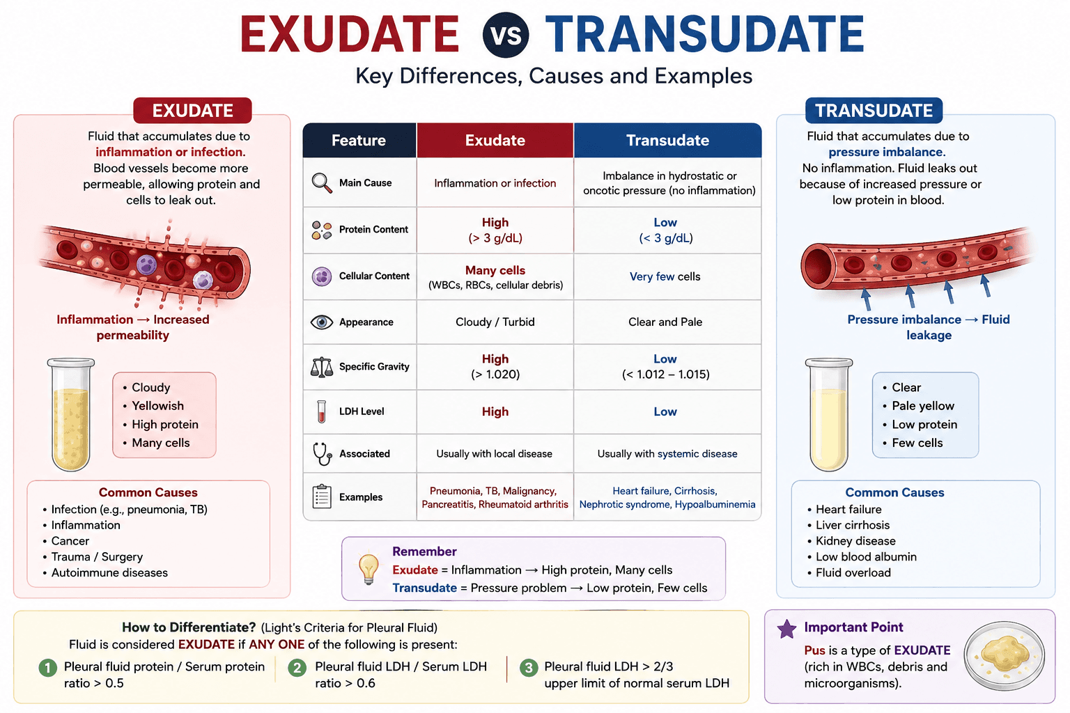
- What is difference between exudate and transudate fluid. — Exudate and Transudate Exudate and transudate are two types of fluid that can accumulate in body tissues or body cavities. They are diffe…
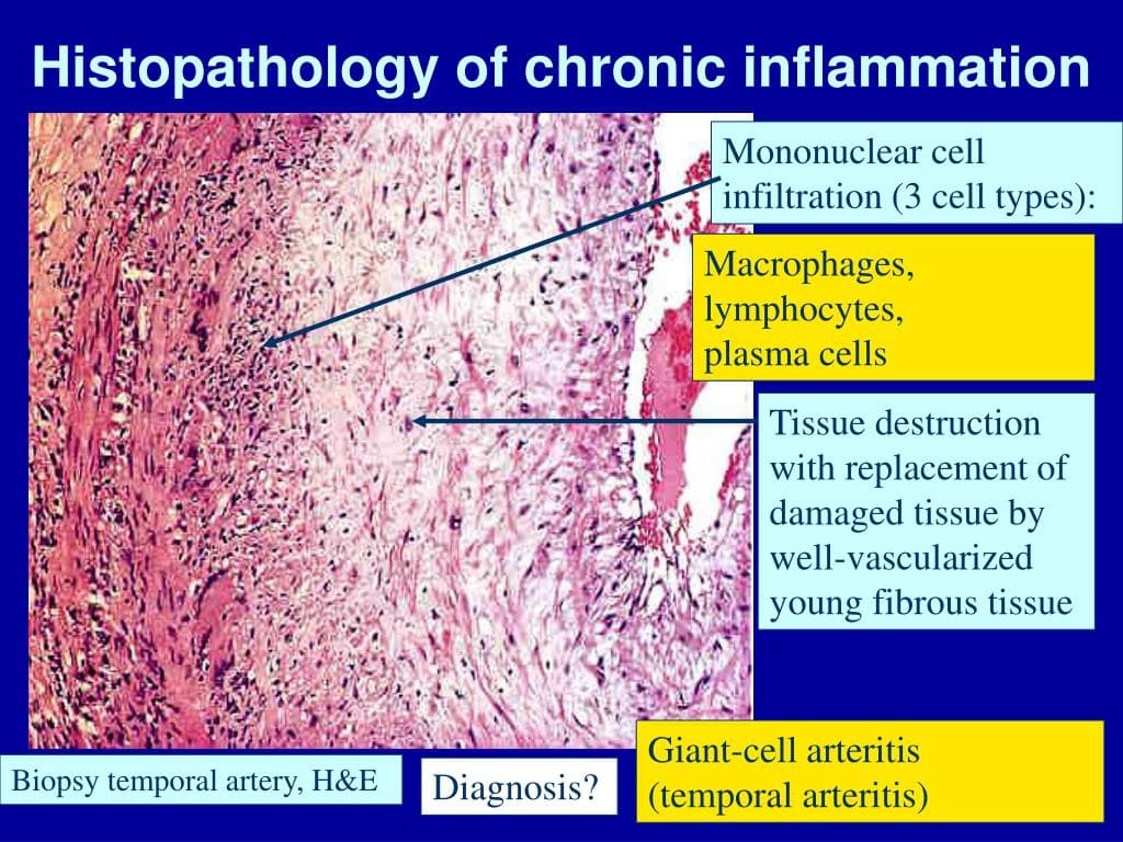
- Chronic Inflammation: Symptoms, Causes, Related Diseases & Treatment — Chronic Inflammation — Notes (Based on the Document) What is Chronic Inflammation? Chronic inflammation is a prolonged inflammatory respo…
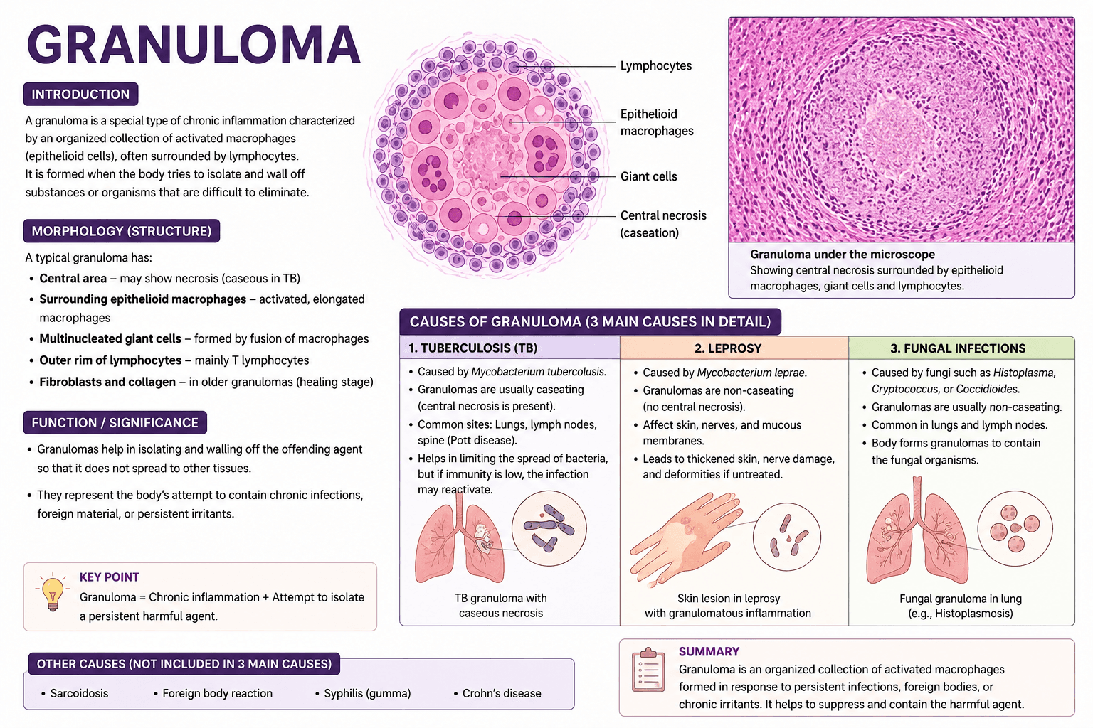
- write a note on granuloma and write down 3 causes of granuloma in details — How Patients Find the Right Doctor Today Finding the right doctor used to mean asking a neighbor, calling your insurance company,…
- Opium Alkaloids and Their Chemical Structures: A Complete Guide — Introduction Opium is of the oldest medicines known to humans, and it is also the starting point for of the most important drug c…
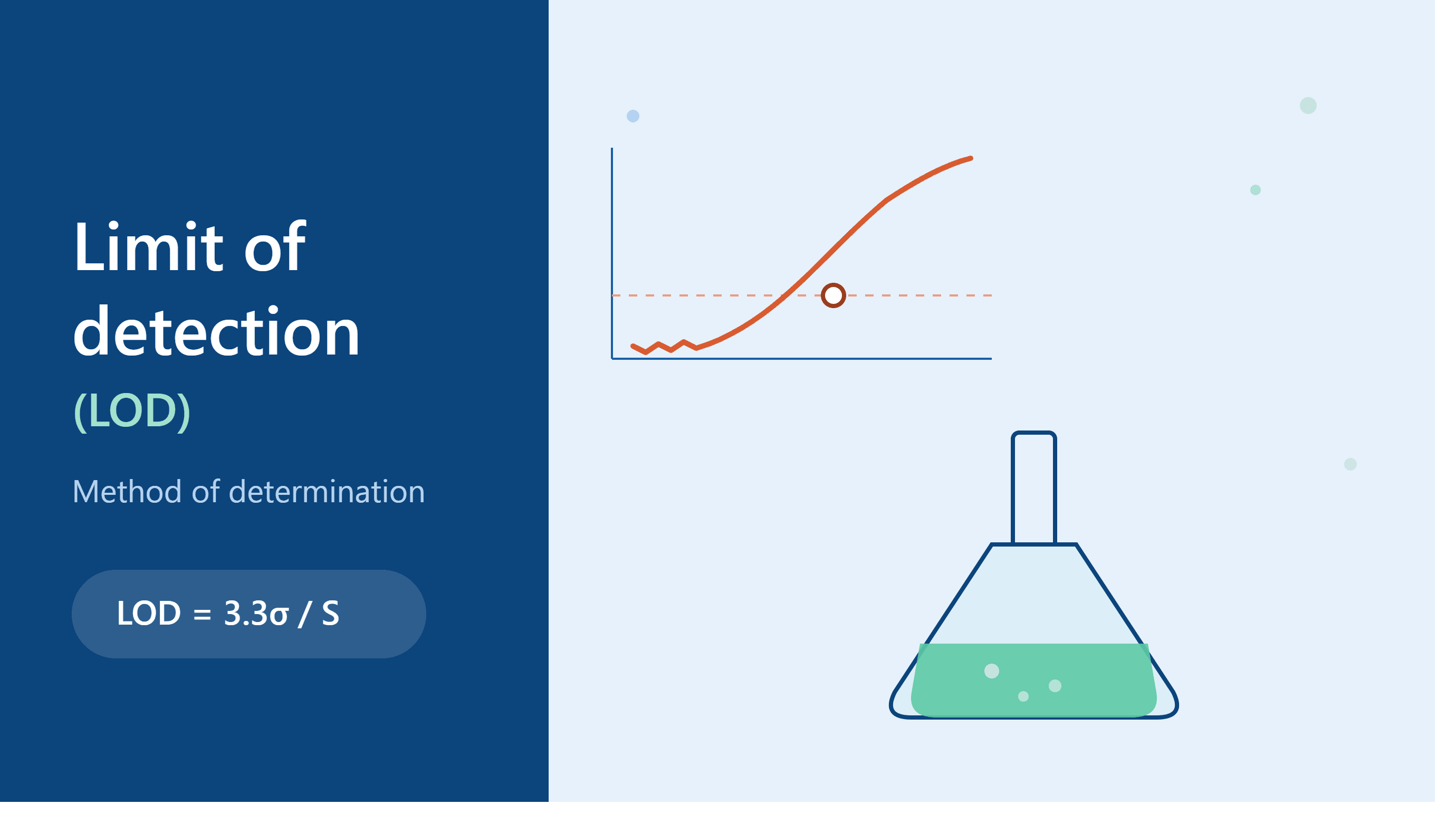
- Limit of Detection (LOD): Method of Determination Explained Simply — How Is the Limit of Detection (LOD) Actually Determined? When an analytical method is validated — an HPLC assay, a UV method, even a simp…
- Understanding Drug–Drug Interactions in Clinical Pharmacy Practice — A practical overview of clinically significant interactions.