Define Hypoxia in Detail: A Pharm-D Study Notes Guide
Understand hypoxia through oxygen delivery, its four major types, diagnostic clues and treatment principles, with practical Pharm-D revision notes.
By Dr. Amina Rahman, PharmD12 min read
Reviewed by Medical Review Team ·
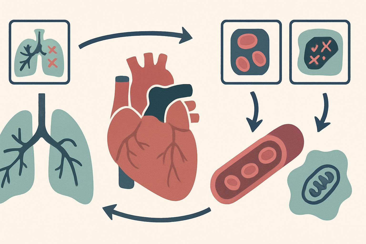
Hypoxia is an inadequate supply of oxygen to tissues, or an inability of cells to use available oxygen, relative to their metabolic needs. It can occur despite normal arterial oxygen measurements when hemoglobin, circulation or cellular oxygen use is impaired.
These Pharm-D notes connect that definition to physiology, investigation and safe treatment. This is educational material, not advertising for any company’s product; compare medicine and device options with a pharmacist.
Quick Answer / Overview
Tissues need functioning lungs, sufficient hemoglobin, adequate blood flow and working cellular machinery to obtain energy from oxygen. Failure at any link can cause hypoxia.
The classic classification includes hypoxemic, anemic, circulatory and histotoxic hypoxia. These mechanisms often overlap in critically ill patients. Hypoxia is a physiological problem, not a single disease, so treatment must address its cause rather than merely improve an oximeter number.
STICKY NOTE — Important point: Hypoxemia describes low oxygen in arterial blood. Hypoxia describes inadequate oxygen availability or use at tissue level. They are related, but not interchangeable.
Key Definitions: Keep the Measurements Separate
- Hypoxemia: abnormally low arterial oxygen tension, usually assessed through PaO₂.
- Anoxia: virtually complete absence of oxygen at tissue level; the extreme end of oxygen deprivation.
- Ischemia: insufficient blood flow to a tissue, restricting oxygen and nutrients while impairing waste removal.
- PaO₂: arterial oxygen partial pressure, reflecting oxygen dissolved in plasma.
- SaO₂: arterial hemoglobin oxygen saturation; direct measurement requires co-oximetry, while some blood gas reports calculate it.
- SpO₂: peripheral oxygen saturation estimated by a pulse oximeter.
- CaO₂: arterial oxygen content, meaning the total oxygen carried in a given volume of arterial blood.
- Dyspnea: the subjective feeling of difficult or uncomfortable breathing.
These terms describe different things. A breathless patient may have normal oxygenation, while someone with significant hypoxemia may report surprisingly little breathlessness.
How Oxygen Reaches Cells
Oxygen delivery depends on content and flow
Most blood oxygen travels attached to hemoglobin. a small amount dissolves in plasma.
A commonly used physiological approximation is:
CaO₂ ≈ (1.34 × hemoglobin × SaO₂) + (0.003 × PaO₂)
Here, hemoglobin is measured in g/dL, SaO₂ is entered as a fraction, PaO₂ in mmHg, and CaO₂ is expressed in mL O₂/dL. Constants vary slightly between references.
Oxygen delivery, DO₂ = cardiac output × CaO₂ × 10, when cardiac output is expressed in L/min.
The implication is practical. A patient with hemoglobin of 7 g/dL carries roughly half as much hemoglobin-bound oxygen as someone with 14 g/dL at the same saturation, assuming comparable hemoglobin function. Their pulse oximeters can show identical readings.
Oxygen extraction provides a reserve
Tissues initially compensate for reduced delivery by extracting more oxygen from blood. delivery becomes critically inadequate, aerobic energy production falls; the threshold varies with metabolic demand and physiological reserve.
Hemoglobin must also release its oxygen. Increased temperature, carbon dioxide, acidity and 2,3-bisphosphoglycerate generally shift the oxygen–hemoglobin dissociation curve rightward, facilitating tissue unloading. Opposite changes shift it leftward.
The curve has a steep lower portion. PaO₂ falls substantially, further small reductions can produce larger saturation drops.
Classification: Four Major Types of Hypoxia
| Type | Main defect | Typical examples | Useful diagnostic clue |
|---|---|---|---|
| Hypoxemic, also called hypoxic | Too little oxygen enters arterial blood | Pneumonia, altitude, hypoventilation | PaO₂ is reduced |
| Anemic | Reduced hemoglobin amount or oxygen-carrying function | Severe anemia, carbon monoxide exposure, methemoglobinemia | PaO₂ may remain normal |
| Circulatory, also called stagnant | Inadequate blood flow delivers insufficient oxygen | Low cardiac output, arterial obstruction | Systemic arterial oxygenation may be preserved |
| Histotoxic | Cells cannot use delivered oxygen effectively | Cyanide poisoning | Tissue extraction may fall despite available oxygen |
Classification is not a bedside verdict. Carbon monoxide poisoning, for example, impairs oxygen carriage and unloading and can also affect cellular function; placing it in teaching category does not describe every mechanism.
Why arterial oxygen can fall
Five mechanisms explain most hypoxemia:
- Low inspired oxygen pressure: occurs at altitude, although atmospheric oxygen percentage remains approximately unchanged.
- Alveolar hypoventilation: insufficient ventilation lowers alveolar oxygen and usually raises carbon dioxide.
- Ventilation–perfusion mismatch: airflow and blood flow are unevenly matched, as in many lung diseases.
- Diffusion limitation: oxygen transfer across the alveolar–capillary membrane is impaired, especially during exertion.
- Right-to-left shunting: blood reaches systemic circulation without adequate contact with ventilated alveoli.
Most mechanisms improve with supplemental oxygen. A substantial true shunt responds less well, so escalating oxygen alone may not solve the underlying problem.
Cellular Response: From Compensation to Injury
Early compensation includes faster breathing, sympathetic activation and increased cardiac output. Local vessels may dilate to improve tissue supply, although hypoxia causes pulmonary arteriolar constriction, redirecting blood away from poorly ventilated lung regions.
Cells then face an energy shortfall. Reduced mitochondrial oxidative phosphorylation lowers adenosine triphosphate, or ATP, compromising membrane pumps and causing sodium, water and calcium disturbances.
Inadequate oxygen delivery or use
 ↓
Reduced mitochondrial ATP production
 ↓
Greater reliance on anaerobic glycolysis
 ↓
Ion-pump failure, cellular swelling and acidosis
 ↓
Persistent injury → membrane damage and cell death
Anaerobic metabolism can increase lactate. However, elevated lactate also occurs through mechanisms other than oxygen deprivation, and a normal value cannot exclude early or localized hypoxia.
Longer-term adaptation involves hypoxia-inducible factors, which regulate genes supporting erythropoietin production, blood-vessel growth and metabolic adjustment. Persistent hypoxemia can produce secondary erythrocytosis and pulmonary hypertension.
The brain and heart are particularly vulnerable. Injury depends on severity, duration, temperature, perfusion and pre-existing disease, not on universal survival time.
Clinical Features and Emergency Recognition
Early findings may include tachypnea, tachycardia, restlessness, headache, reduced concentration and breathlessness. Severe or prolonged hypoxia can cause confusion, exhaustion, arrhythmias, impaired consciousness and organ dysfunction.
Cyanosis is an unreliable screening sign. Its visibility depends partly on the absolute amount of deoxygenated hemoglobin, skin characteristics and perfusion; severe anemia can mask it despite dangerous oxygen deficiency.
Call emergency services for severe breathing difficulty, inability to speak normally because of breathlessness, new confusion, collapse, chest pain or blue-gray lips or tongue. Do not wait for an oximeter reading.
For most adults at sea level without a clinician-set lower baseline, a persistent resting SpO₂ of 92% or less warrants urgent assessment; 90% or less generally requires emergency evaluation. Symptoms override thresholds, and altitude, age and established disease modify interpretation.
IMPORTANT — Safety: A normal-looking saturation does not exclude carbon monoxide poisoning, severe anemia or impaired tissue perfusion. Treat the clinical situation, not just the display.
Assessment: What Each Test Can and Cannot Tell You
Pulse oximetry
Check technique first. Let the person rest, warm a cold hand, minimize movement and wait for a stable signal; remove nail products if they interfere.
Poor perfusion, movement, device quality and skin pigmentation can affect accuracy. Some devices may overestimate oxygenation in people with darker skin, particularly at lower saturations. Consumer wellness devices are not equivalent to validated medical equipment.
Conventional pulse oximeters cannot reliably distinguish oxyhemoglobin from carboxyhemoglobin. Methemoglobinemia also makes their readings misleading.
Arterial blood gas and co-oximetry
An arterial blood gas, or ABG, evaluates PaO₂, carbon dioxide tension and acid–base status. Interpret it alongside the oxygen device, delivered concentration and sampling conditions.
A venous blood gas can help assess acid–base disturbances in selected settings, but venous oxygen tension cannot replace PaO₂ for assessing arterial oxygenation.
Co-oximetry measures different hemoglobin species, including carboxyhemoglobin and methemoglobin. Request it when exposure history, medicine use or a mismatch between clinical findings and routine readings raises suspicion.
Finding the missing link
A complete blood count checks hemoglobin; examination, electrocardiography, imaging and perfusion assessment address circulation and cardiopulmonary causes. Lactate trends may help but remain nonspecific.
The alveolar–arterial oxygen gradient compares estimated alveolar oxygen with measured PaO₂. It is often normal in pure hypoventilation or low inspired oxygen pressure and increased with ventilation–perfusion mismatch, diffusion impairment or shunting. Age and inspired oxygen affect interpretation.
A useful examination question is: Which variable is abnormal—pressure, saturation, content, flow or utilization? This approach is more informative than asking whether oxygen is “low.”
Treatment Principles: Oxygen Is Support, Not the Whole Treatment
Start with airway, breathing and circulation. Serious hypoxia requires emergency assessment, monitoring and prompt treatment of the underlying cause; investigations should not unnecessarily delay stabilization.
Supplemental oxygen is generally indicated for clinically important hypoxemia. Delivery equipment and flow depend on severity, the target saturation and whether ventilatory support is needed. Oxygen alone does not correct inadequate ventilation or remove accumulated carbon dioxide.
British Thoracic Society guidance commonly uses targets of 94–98% for most acutely ill adults and 88–92% for those at risk of hypercapnic respiratory failure, pending blood gas results. Local protocols and individual prescriptions take precedence; these are not universal targets for every condition or age group.
Do not withhold lifesaving oxygen because a patient has chronic obstructive pulmonary disease. Give controlled oxygen, obtain blood gases and reassess promptly.
Cause-specific care may include bronchodilators for bronchospasm, antimicrobial treatment for appropriate infections, correction of significant anemia or restoration of perfusion. Suspected carbon monoxide or cyanide poisoning requires urgent specialist assessment and exposure-specific treatment.
Oxygen has risks. Excess exposure can contribute to hypercapnia in susceptible patients, absorption atelectasis and oxidative injury; oxygen equipment also creates a major fire hazard. Keep it away from smoking, flames and petroleum-based products.
There is no single medicine or fixed dose for “hypoxia.” Confirm every medicine dose and prescribed oxygen setting with the treating doctor or pharmacist; do not adjust treatment from these notes.
Pharmacy Practice: Medication Clues and Counseling
A medicine history can reveal the mechanism. Opioids such as morphine and benzodiazepines such as diazepam can suppress ventilation, particularly when combined with alcohol or other sedatives. Unexpected sleepiness with slow breathing is an emergency, not simply a tolerable adverse effect.
Dapsone, an antimicrobial, and benzocaine, a local anesthetic, can cause methemoglobinemia. Ask about recent dental or throat products, not prescription tablets. If a nonessential topical product is suspected, stop further use and seek urgent assessment; severe symptoms require emergency care.
Anticoagulants and nonsteroidal anti-inflammatory drugs can contribute to bleeding and subsequent anemia. New pallor, faintness, black stools or vomiting blood needs prompt evaluation even when SpO₂ appears normal.
Check active ingredients, strengths and duplicate sedating ingredients on labels. Generic names improve communication across brands, but formulations and release characteristics are not automatically interchangeable.
REMEMBER — The missing-number check: Saturation tells you how full the available hemoglobin binding sites are. It does not tell you how much hemoglobin exists or how much blood reaches the tissue.
Quick Pharm D Notes
- Definition: tissue oxygen supply or utilization is insufficient for metabolic demand.
- Classification: hypoxemic, anemic, circulatory and histotoxic.
- Process: impaired oxygen delivery or use reduces oxidative phosphorylation and ATP availability.
- Examples: pneumonia, severe anemia, arterial obstruction and cyanide toxicity illustrate different failure points.
- Clinical significance: persistent hypoxia can cause organ injury despite initially successful compensation.
- Exam point: normal PaO₂ does not guarantee normal oxygen content or tissue oxygenation.
- Interpretation point: document oxygen support whenever interpreting blood gases or saturation trends.
Common Mistakes and Important Differences
| Common assumption | Correct interpretation |
|---|---|
| Hypoxia and ischemia mean the same thing | Ischemia reduces blood flow; hypoxia can occur with preserved flow |
| Every breathless patient needs oxygen | Breathlessness has oxygen-related and non-oxygen-related causes |
| Normal PaO₂ excludes oxygen-related toxicity | Dyshemoglobinemia may leave dissolved oxygen relatively unchanged |
| More oxygen is always safer | Excess oxygen can harm; treatment needs a target and reassessment |
Avoid a second trap: assuming oxygen correction proves recovery. An improved saturation may coexist with respiratory fatigue, rising carbon dioxide or deteriorating circulation, so reassess mental status, breathing effort and perfusion.
Frequently Asked Questions
Can hypoxia occur with a normal pulse oximeter reading?
Yes. Severe anemia, poor circulation, carbon monoxide exposure and impaired cellular oxygen use can cause tissue hypoxia despite reassuring conventional readings.
What is the difference between hypoxia and hypoxemia?
Hypoxemia means low arterial oxygen tension. Hypoxia means inadequate tissue oxygen availability or utilization; hypoxemia is possible cause.
Is hypoxia always accompanied by blue lips?
No. Cyanosis may be absent, difficult to recognize or delayed. Do not use lip color alone to decide whether someone needs emergency help.
Can oxygen treat every type of hypoxia?
No. It supports oxygenation, but cannot independently correct severe anemia, an obstructed artery or blocked cellular oxygen utilization. Some poisonings still require oxygen plus specialist treatment.
Can medicines cause hypoxia?
Yes. Mechanisms include respiratory depression, abnormal hemoglobin formation, bleeding-related anemia and drug-induced lung injury. The medicine history should include nonprescription products and recreational substances.
Can hypoxia cause permanent brain damage?
Yes, particularly when severe or prolonged. Outcome depends on the cause, duration, circulation and speed of effective treatment; saturation reading cannot predict it.
Last-Minute Revision
- Separate oxygen pressure, saturation, content, delivery and utilization.
- Classify the failed step before selecting investigations.
- Expect overlapping mechanisms in complex illness.
- Recognize that cyanosis and pulse oximetry can mislead.
- Use co-oximetry when abnormal hemoglobin species are suspected.
- Treat the cause while supporting oxygenation and ventilation.
References
- Guyton and Hall Textbook of Medical Physiology: oxygen transport and respiratory physiology.
- Robbins & Cotran Pathologic Basis of Disease: cellular injury and adaptation.
- British Thoracic Society: Guideline for Oxygen Use in Adults in Healthcare and Emergency Settings.
- U.S. Food and Drug Administration: Pulse Oximeters: Technology, Accuracy, and Limitations.
Important Medical Disclaimer
This article is for general educational purposes and is not a substitute for advice from a qualified doctor or pharmacist. Always consult your doctor/pharmacist before using any medicine, changing a dose, or starting treatment.
Related reading
If this article on Define Hypoxia in Detail: A Pharm-D Study Notes Guide was helpful, explore these related guides for more practical information on similar topics.
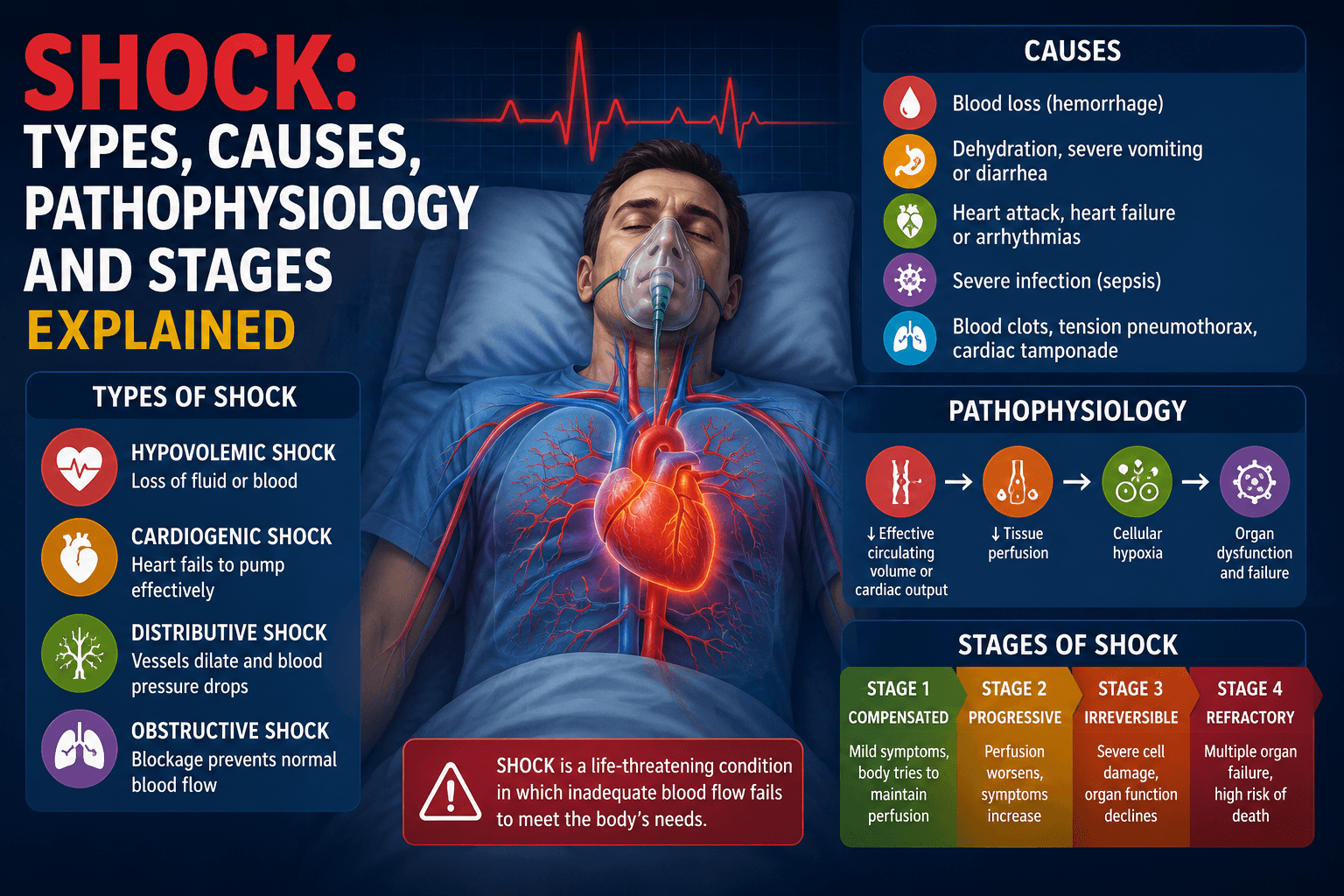
- Shock: Types, Causes, Pathophysiology and Stages Explained — Shock: Types, Causes, Pathophysiology and Stages Explained (Complete Pharm D Notes) Introduction Shock is of the most important topic…
- Define Hemorrhage: Detailed Pharm-D Notes and Safety — Understand hemorrhage through classification, mechanisms, morphology, clinical signs, and medicine-related risks, with clear exam notes a…
- Apoptosis Defined: Mechanisms and Pharm-D Study Notes — Define apoptosis with clear pathways, morphology, clinical examples and exam distinctions, plus practical safety notes on apoptosis-targe…
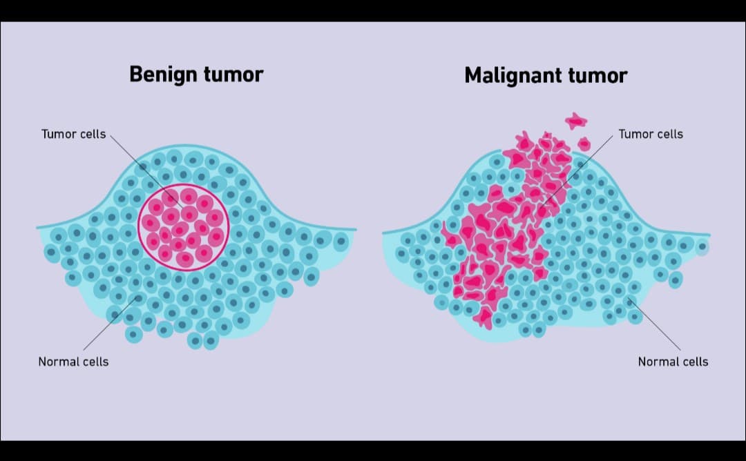
- Define neoplasia. Discuss in detail the features of malignant tumors. — Neoplasia — Definition Neoplasia is an abnormal, uncontrolled and excessive proliferation of cells that continues even after the original…
- Know the causes of white hair and easy ways to prevent it naturally — Learn why hair turns white, which natural habits support healthy growth, and when early greying needs medical checks rather than another…
- Define acute inflammation describe different morphological patterns of acute inflammation — Definition Acute inflammation is a rapid and short-duration inflammatory response of vascularized tissue to injury or infection. It is ch…