Define Hemorrhage: Detailed Pharm-D Notes and Safety
Understand hemorrhage through classification, mechanisms, morphology, clinical signs, and medicine-related risks, with clear exam notes and safety advice.
By Dr. Amina Rahman, PharmD13 min read
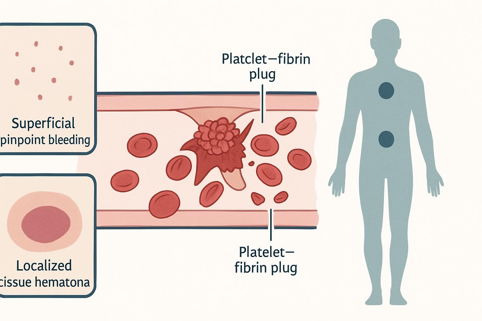
Hemorrhage is the escape of blood from the heart or blood vessels into surrounding tissues, body cavities, or outside the body. Its clinical importance depends on the amount, speed, and location of bleeding, together with the patient’s ability to compensate and form an effective clot.
These Pharm-D study notes connect pathology with bedside assessment and medication safety. They are educational only, not advertising for any company’s product; compare medicine options with a pharmacist rather than choosing by brand.
Quick Answer / Overview
Hemorrhage means bleeding. It may follow an obvious injury, occur inside an organ without visible blood, or develop because platelets or clotting factors cannot maintain hemostasis—the process that stops bleeding.
A small intracranial hemorrhage can be more dangerous than a larger superficial bruise because the skull cannot accommodate expansion without threatening brain function. Conversely, slow, unnoticed gastrointestinal bleeding may present mainly as iron-deficiency anemia.
Location changes the risk. Visible blood alone does not measure severity.
Key Definitions
- Hemostasis: The coordinated vascular, platelet, and coagulation responses that limit blood loss.
- Hematoma: A localized collection of blood within tissue, often producing swelling.
- Thrombocytopenia: A platelet count below the laboratory reference range.
- Coagulopathy: Impaired clot formation caused by abnormalities of coagulation, often involving clotting factors.
- Occult bleeding: Blood loss that is not apparent on ordinary inspection.
- Hemorrhagic shock: Inadequate tissue perfusion caused by sufficiently severe blood loss.
Hemorrhage and shock are not interchangeable. Bleeding may occur without shock, whereas shock can arise from causes unrelated to blood loss.
Classification: Describe More Than Feature
Several classifications apply simultaneously. A patient can have acute, internal, arterial hemorrhage from a ruptured vessel; these terms describe different aspects of the same event.
| Classification basis | Categories | Practical meaning |
|---|---|---|
| Visibility or destination | External, internal, externalized | Blood leaves through a wound, remains inside, or exits through a natural opening |
| Vessel involved | Arterial, venous, capillary | Identifies the vascular source, not a reliable bedside severity score |
| Time course | Acute or chronic | Rapid loss threatens circulation; sustained loss may deplete iron |
| Mechanism | Vessel rupture or microscopic leakage | Distinguishes structural disruption from leakage through small-vessel walls |
| Context | Traumatic, procedural, spontaneous | Directs the search for injury, treatment complications, or disease |
Arterial bleeding may be pulsatile, venous bleeding may flow steadily, and capillary bleeding often oozes. These are teaching patterns, not dependable rules: lighting, oxygenation, wound depth, and mixed vessel injury alter appearances.
Timing after injury or surgery
Traditional surgical terminology separates primary hemorrhage, occurring at injury or operation, from reactionary hemorrhage, recurring relatively soon afterward as pressure returns or hemostasis fails. Secondary hemorrhage develops later, often with infection, tissue breakdown, or vessel erosion.
Exact time windows vary across teaching sources. Explain the underlying event rather than memorizing a rigid interval as universally applicable.
Causes and Mechanisms of Hemorrhage
Normal hemostasis needs an intact vessel wall, functioning platelets, adequate coagulation factors, and controlled fibrinolysis—the breakdown of fibrin. Failure at any point can permit bleeding, particularly when several problems coexist.
Vessel damage
Trauma, surgery, ulcers, tumors, aneurysm rupture, and inflamed vessels can disrupt vascular integrity. Hypertension contributes to some intracerebral hemorrhages, while portal hypertension can produce gastroesophageal varices that may rupture.
The lesion matters. Correcting a coagulation abnormality does not close an actively bleeding ulcer or repair a torn artery.
Platelet and vessel-wall disorders
Low platelet counts, inherited platelet defects, antiplatelet medicines, and platelet dysfunction in advanced kidney disease commonly produce mucosal bleeding, petechiae, or easy bruising. Vitamin C deficiency can weaken supporting connective tissue and increase vascular fragility.
In classical pathology, hemorrhage by rhexis means bleeding through a ruptured vessel; hemorrhage by diapedesis describes red-cell passage through small-vessel walls without a grossly visible tear. Diapedesis here does not mean ordinary leukocyte migration during inflammation.
Coagulation and fibrinolytic disorders
Hemophilia, vitamin K deficiency, severe liver disease, and anticoagulants can impair fibrin formation. Excessive fibrinolysis can destabilize a clot after it forms.
Disseminated intravascular coagulation, or DIC, produces widespread coagulation activation with consumption of platelets and factors. Bleeding and thrombosis can therefore occur together.
STICKY NOTE — Mechanism: Classify the cause as vessel, platelet, coagulation-factor, or fibrinolytic dysfunction. Then ask whether multiple mechanisms are operating.
Morphological Patterns and Named Bleeding Sites
Size labels are useful shorthand. Their boundaries are approximate and vary slightly among pathology and dermatology references; distribution, palpability, associated symptoms, and progression usually matter more clinically.
| Pattern | Typical description | Interpretation |
|---|---|---|
| Petechiae | Pinpoint, non-blanching spots, usually around 1–2 mm | Suggest platelet or small-vessel problems, although causes vary |
| Purpura | Larger non-blanching lesions; also an umbrella term for cutaneous bleeding | Palpable purpura raises concern for small-vessel vasculitis |
| Ecchymosis | A larger, flat bruise, commonly over 1 cm | May follow trauma or impaired hemostasis |
| Hematoma | A localized blood collection in tissue | Can compress nerves, vessels, or adjacent structures |
Bruises change color as hemoglobin breaks down and macrophages clear blood products. However, color cannot reliably date an injury; depth, skin tone, location, and individual healing affect the appearance.
Bleeding into the pleural cavity is hemothorax; into the pericardial sac, hemopericardium; into the peritoneal cavity, hemoperitoneum; and into a joint, hemarthrosis. These names identify location, not cause.
Hematemesis means vomiting blood. Melena usually means black, tarry stool containing digested blood, commonly from an upper gastrointestinal source. Hematochezia is red or maroon blood passed rectally, usually from lower gastrointestinal bleeding, although brisk upper gastrointestinal bleeding can also cause it.
Hemoptysis means coughing blood from the respiratory tract; hematuria means blood in urine. Swallowed nasal blood can imitate gastrointestinal bleeding, so establish how the blood first appeared.
Physiological Consequences: Rate, Volume, and Site
Rapid blood loss reduces venous return and cardiac filling. Stroke volume falls, sympathetic compensation increases, and peripheral vasoconstriction attempts to preserve blood flow to vital organs.
Vessel injury or failed hemostasis
 ↓
Continued blood loss
 ↓
Reduced circulating volume and oxygen-carrying capacity
 ↓
Compensation: faster pulse and peripheral vasoconstriction
 ↓
If bleeding exceeds compensation: tissue hypoperfusion
 ↓
Organ dysfunction and possible hemorrhagic shock
Compensation can hide deterioration. Blood pressure may remain near normal early, and beta-blockers or other patient factors can blunt the expected tachycardia.
Acute whole-blood loss may initially leave hemoglobin concentration deceptively normal because plasma and red cells are lost together. The measured concentration commonly falls after fluid redistribution or resuscitation; serial results and the clinical picture matter more than early sample.
Chronic external blood loss removes iron from the body and may cause microcytic, hypochromic anemia—small, pale red cells. Iron depletion can precede those characteristic red-cell changes.
Local effects can dominate: intracranial blood raises pressure, hemopericardium can cause cardiac tamponade, and an expanding limb hematoma can contribute to compartment syndrome. A collection need not be enormous to threaten an organ.
REMEMBER — Laboratory trap: A normal initial hemoglobin does not exclude major acute hemorrhage. Do not wait for anemia before recognizing instability.
Recognition and Investigations
First assess airway, breathing, circulation, mental status, and blood loss. Severe bleeding is an emergency; investigations should support stabilization and source control rather than postpone them.
Urgent warning signs include uncontrolled bleeding, fainting, confusion, cold clammy skin, breathlessness, vomiting blood, or black tarry stools. Sudden severe headache, new weakness, or reduced consciousness can indicate intracranial hemorrhage, even without visible blood.
Clinical assessment should establish the injury or procedure history, bleeding duration, menstrual and gastrointestinal symptoms, family history, and all medicines. Ask about nonprescription products explicitly; patients may not consider painkillers or herbal preparations to be medicines.
Common investigations include:
- Complete blood count: Hemoglobin trend, red-cell indices, and platelet count.
- Blood film: Selected clues to platelet or red-cell abnormalities.
- Prothrombin time and international normalized ratio: Assessment of parts of coagulation; INR is particularly used for warfarin monitoring.
- Activated partial thromboplastin time: Assessment of other coagulation pathways and selected treatment effects.
- Fibrinogen and D-dimer: Useful in appropriate contexts, including suspected DIC; neither should be interpreted alone.
- Renal and liver function: Potential contributors to bleeding and altered medicine clearance.
- Blood grouping and compatibility testing: When transfusion may be needed.
Imaging, endoscopy, or targeted procedures identify the source. Pregnancy-related bleeding needs obstetric assessment when relevant.
Routine coagulation tests have limits. Normal results do not exclude platelet dysfunction, von Willebrand disease, or a clinically relevant direct oral anticoagulant effect; interpretation depends on the drug, assay, and time since dosing.
Important Differences Students Commonly Confuse
Patterns guide investigation, but they are not diagnoses. Mixed disorders and severe disease can blur the distinctions below.
| Feature | Platelet-type bleeding | Coagulation-factor-type bleeding |
|---|---|---|
| Common sites | Skin and mucous membranes | Deep tissues, muscles, and joints |
| Typical findings | Petechiae, gum bleeding, nosebleeds | Large hematomas, hemarthrosis |
| After procedures | Often immediate oozing | May include delayed or recurrent bleeding |
| Platelet count | Low or normal, depending on cause | Often normal in isolated factor disorders |
Von Willebrand disease is a useful exception to simplistic rules: it affects platelet adhesion and may reduce factor VIII levels, yet often presents with mucosal bleeding.
Also distinguish hemorrhage from hemolysis, the destruction of red cells. Both can produce anemia, but the investigations and treatment differ.
Pharmacy Practice: Medicines, Reversal, and Counseling
Medication review can change management. Record the exact product, strength, indication, last dose, renal function, and recent additions rather than simply documenting that the patient takes a “blood thinner.”
Medicines that increase bleeding risk
Warfarin, a vitamin K antagonist; apixaban and rivaroxaban, direct factor Xa inhibitors; and dabigatran, a direct thrombin inhibitor, impair coagulation through different mechanisms. Aspirin and clopidogrel inhibit platelet function.
Ibuprofen and naproxen, nonsteroidal anti-inflammatory drugs, can damage gastrointestinal mucosa and affect platelet function. Combining an anticoagulant, an antiplatelet medicine, and an NSAID may substantially increase bleeding risk, particularly with additional patient risk factors.
Check duplicate ingredients. In settings where painkillers are readily available without prescription, combination headache or cold products can quietly add aspirin or an NSAID to an existing regimen.
Treatment principles and safety limits
Control the bleeding source first or alongside resuscitation. Depending on the cause, clinicians may use direct compression, surgery, endoscopy, interventional radiology, blood components, or targeted reversal.
Reversal is drug-specific: vitamin K with appropriate factor replacement is used in selected serious warfarin-associated bleeding; idarucizumab reverses dabigatran. Factor Xa inhibitor reversal strategies depend on local authorization, availability, severity, and protocols. Protamine reverses unfractionated heparin and partly reverses low-molecular-weight heparin.
Tranexamic acid, an antifibrinolytic, reduces fibrin breakdown and has specific evidence-based uses, including selected trauma and obstetric bleeding. It is not a universal treatment for every hemorrhage or a substitute for source control.
Potential harms matter too. Reversal and prohemostatic treatment can increase thrombotic risk; transfusion can cause reactions or circulatory overload, and tranexamic acid requires indication-specific precautions, including renal assessment.
No universal hemorrhage dose exists. Treatment depends on indication, severity, timing, weight, laboratory findings, and organ function. Confirm every medicine dose with the treating doctor or pharmacist and the locally approved label or protocol.
IMPORTANT — Do not self-adjust: Minor bruising does not automatically justify stopping a prescribed anticoagulant. Major or uncontrolled bleeding needs emergency assessment; the emergency team should direct withholding and reversal.
Practical First Aid and Common Mistakes
For external bleeding, apply firm, continuous pressure with a clean dressing. If it becomes soaked, keep pressure on and add material rather than repeatedly lifting it to inspect the wound.
Call emergency services for severe, uncontrolled, or suspected internal bleeding. Do not remove an embedded object; press around it. Life-threatening limb bleeding may require a tourniquet, preferably applied by someone trained or following emergency-dispatch instructions.
For a straightforward nosebleed, sit leaning forward and pinch the soft nose continuously. Do not tilt the head backward. Persistent or heavy bleeding, dizziness, injury, or anticoagulant use warrants prompt assessment.
Avoid these common errors:
- Estimating severity solely from the visible blood pool.
- Assuming normal blood pressure excludes dangerous bleeding.
- Treating anemia with iron without investigating blood loss.
- Attributing hematuria to anticoagulants without assessing urinary disease.
- Calling every dark stool melena: iron and bismuth can darken stool, but tarry stool or systemic symptoms still require assessment.
Quick Pharm D Notes
- Definition: Blood escapes from the cardiovascular system.
- Classification: Describe location, source, rate, mechanism, and context.
- Process: Vascular disruption or ineffective hemostasis permits continued blood loss.
- Examples: Petechiae, ecchymosis, hematoma, hemothorax, and hemarthrosis.
- Clinical significance: Consider hypoperfusion, anemia, compression, and functional impairment.
- Exam point: Explain why a small critical-site bleed can outweigh a larger superficial one.
Frequently Asked Questions
Is hemorrhage always visible?
No. Blood may accumulate in the abdomen, chest, brain, or deep tissues. Pain, swelling, neurological changes, or circulatory deterioration may appear before any external blood is noticed.
Does every hemorrhage cause shock?
No. Shock occurs when blood loss overwhelms compensation and compromises tissue perfusion. Small superficial bleeds usually do not cause it.
Can hemorrhage occur with normal platelets?
Yes. Platelet dysfunction, coagulation-factor deficiency, vessel injury, and anticoagulant treatment can cause bleeding despite a normal platelet count.
Are petechiae harmless?
Not necessarily. Local pressure can cause them, but new widespread petechiae, fever, illness, or mucosal bleeding needs prompt assessment. A non-blanching rash with severe illness is an emergency.
Should aspirin be stopped when bruises appear?
Do not stop prescribed aspirin solely because of minor bruising without advice, particularly after a coronary stent. Report new or worsening bruising; severe bleeding requires emergency care.
Can bleeding recur after it appears to stop?
Yes. Clot disruption, an untreated lesion, infection, or persisting anticoagulant effects may cause rebleeding. Follow the observation and return instructions given after treatment.
Last-Minute Revision
- Hemorrhage describes escaped blood, not its cause.
- Rate, volume, site, and patient reserve determine consequences.
- Early hemoglobin and blood pressure can mislead.
- Platelet disorders commonly cause mucocutaneous bleeding.
- Factor disorders commonly cause deeper bleeding.
- Source control and medication review belong together.
References
- Robbins & Cotran Pathologic Basis of Disease: hemodynamic disorders and hemorrhage.
- Harrison’s Principles of Internal Medicine: bleeding and thrombotic disorders.
- MSD Manual Professional Edition: “Overview of Hemostasis” and “Excessive Bleeding.”
- American College of Cardiology: 2020 ACC Expert Consensus Decision Pathway on Management of Bleeding in Patients on Oral Anticoagulants.
- World Health Organization: WHO recommendation on tranexamic acid for the treatment of postpartum haemorrhage.
Important Medical Disclaimer
This article is for general educational purposes and is not a substitute for advice from a qualified doctor or pharmacist. Always consult your doctor/pharmacist before using any medicine, changing a dose, or starting treatment.
Related reading
If this article on Define Hemorrhage: Detailed Pharm-D Notes and Safety was helpful, explore these related guides for more practical information on similar topics.
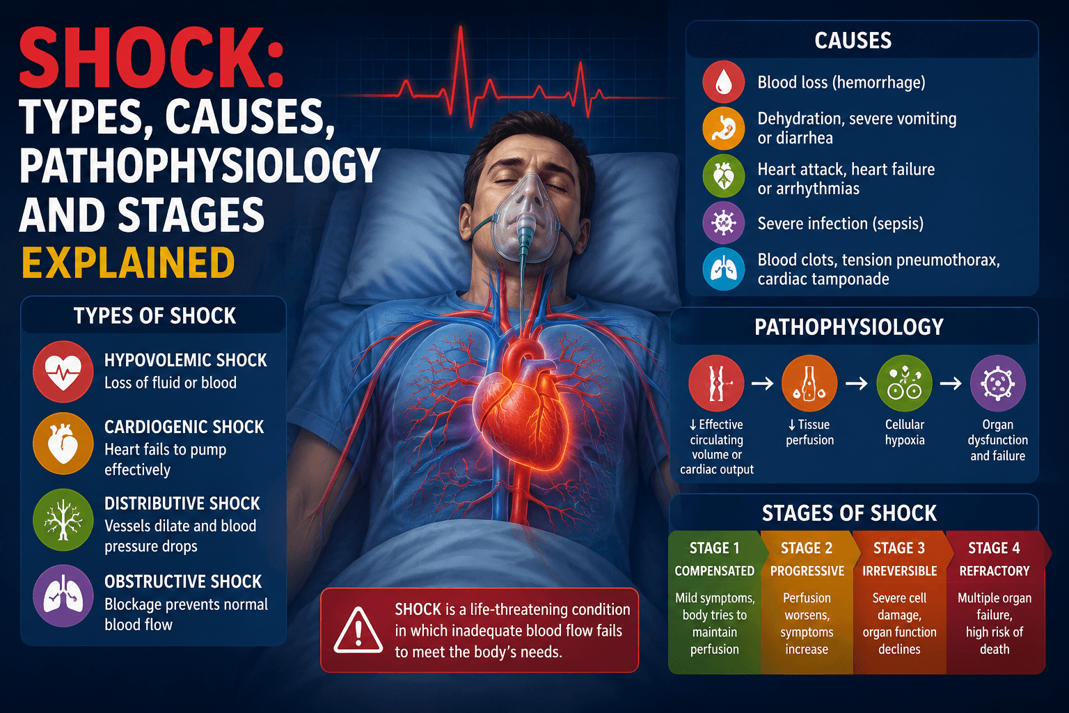
- Shock: Types, Causes, Pathophysiology and Stages Explained — Shock: Types, Causes, Pathophysiology and Stages Explained (Complete Pharm D Notes) Introduction Shock is of the most important topic…
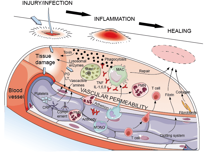
- Define acute inflammation describe different morphological patterns of acute inflammation — Definition Acute inflammation is a rapid and short-duration inflammatory response of vascularized tissue to injury or infection. It is ch…
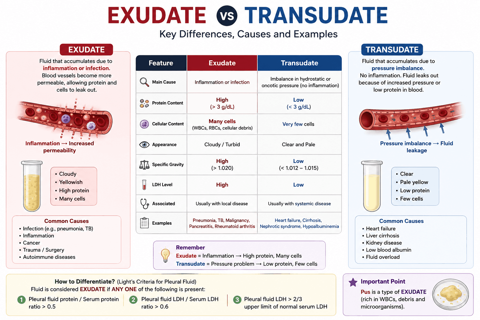
- What is difference between exudate and transudate fluid. — Exudate and Transudate Exudate and transudate are two types of fluid that can accumulate in body tissues or body cavities. They are diffe…
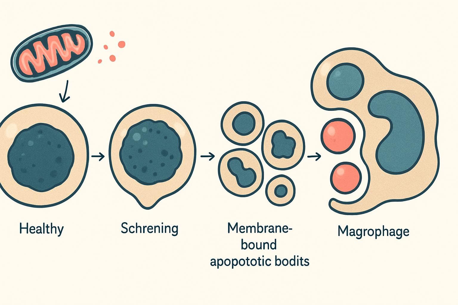
- Apoptosis Defined: Mechanisms and Pharm-D Study Notes — Define apoptosis with clear pathways, morphology, clinical examples and exam distinctions, plus practical safety notes on apoptosis-targe…
- Adverse Drug Reaction Reporting Systems — Pharmacovigilance fundamentals for students and practicing pharmacists.
- Ascard Tablet Overview: Uses, Dosage and Safe Use Tips — Understand what Ascard contains, why doctors prescribe it, and how to avoid bleeding risks, missed-dose mistakes and unsafe tablet substi…